12.1 Basic Structure and Function of the Nervous System
Learning objectives.
By the end of this section, you will be able to:
- Identify the anatomical and functional divisions of the nervous system
- Relate the functional and structural differences between gray matter and white matter structures of the nervous system to the structure of neurons
- List the basic functions of the nervous system
The picture you have in your mind of the nervous system probably includes the brain , the nervous tissue contained within the cranium, and the spinal cord , the extension of nervous tissue within the vertebral column. That suggests it is made of two organs—and you may not even think of the spinal cord as an organ—but the nervous system is a very complex structure. Within the brain, many different and separate regions are responsible for many different and separate functions. It is as if the nervous system is composed of many organs that all look similar and can only be differentiated using tools such as the microscope or electrophysiology. In comparison, it is easy to see that the stomach is different than the esophagus or the liver, so you can imagine the digestive system as a collection of specific organs.

The Central and Peripheral Nervous Systems
The nervous system can be divided into two major regions: the central and peripheral nervous systems. The central nervous system (CNS) is the brain and spinal cord, and the peripheral nervous system (PNS) is everything else ( Figure 12.2 ). The brain is contained within the cranial cavity of the skull, and the spinal cord is contained within the vertebral cavity of the vertebral column. It is a bit of an oversimplification to say that the CNS is what is inside these two cavities and the peripheral nervous system is outside of them, but that is one way to start to think about it. In actuality, there are some elements of the peripheral nervous system that are within the cranial or vertebral cavities. The peripheral nervous system is so named because it is on the periphery—meaning beyond the brain and spinal cord. Depending on different aspects of the nervous system, the dividing line between central and peripheral is not necessarily universal.
Nervous tissue, present in both the CNS and PNS, contains two basic types of cells: neurons and glial cells. A glial cell is one of a variety of cells that provide a framework of tissue that supports the neurons and their activities. The neuron is the more functionally important of the two, in terms of the communicative function of the nervous system. To describe the functional divisions of the nervous system, it is important to understand the structure of a neuron. Neurons are cells and therefore have a soma , or cell body, but they also have extensions of the cell; each extension is generally referred to as a process . There is one important process that every neuron has called an axon , which is the fiber that connects a neuron with its target. Another type of process that branches off from the soma is the dendrite . Dendrites are responsible for receiving most of the input from other neurons. Looking at nervous tissue, there are regions that predominantly contain cell bodies and regions that are largely composed of just axons. These two regions within nervous system structures are often referred to as gray matter (the regions with many cell bodies and dendrites) or white matter (the regions with many axons). Figure 12.3 demonstrates the appearance of these regions in the brain and spinal cord. The colors ascribed to these regions are what would be seen in “fresh,” or unstained, nervous tissue. Gray matter is not necessarily gray. It can be pinkish because of blood content, or even slightly tan, depending on how long the tissue has been preserved. But white matter is white because axons are insulated by a lipid-rich substance called myelin . Lipids can appear as white (“fatty”) material, much like the fat on a raw piece of chicken or beef. Actually, gray matter may have that color ascribed to it because next to the white matter, it is just darker—hence, gray.
The distinction between gray matter and white matter is most often applied to central nervous tissue, which has large regions that can be seen with the unaided eye. When looking at peripheral structures, often a microscope is used and the tissue is stained with artificial colors. That is not to say that central nervous tissue cannot be stained and viewed under a microscope, but unstained tissue is most likely from the CNS—for example, a frontal section of the brain or cross section of the spinal cord.
Regardless of the appearance of stained or unstained tissue, the cell bodies of neurons or axons can be located in discrete anatomical structures that need to be named. Those names are specific to whether the structure is central or peripheral. A localized collection of neuron cell bodies in the CNS is referred to as a nucleus . In the PNS, a cluster of neuron cell bodies is referred to as a ganglion . Figure 12.4 indicates how the term nucleus has a few different meanings within anatomy and physiology. It is the center of an atom, where protons and neutrons are found; it is the center of a cell, where the DNA is found; and it is a center of some function in the CNS. There is also a potentially confusing use of the word ganglion (plural = ganglia) that has a historical explanation. In the central nervous system, there is a group of nuclei that are connected together and were once called the basal ganglia before “ganglion” became accepted as a description for a peripheral structure. Some sources refer to this group of nuclei as the “basal nuclei” to avoid confusion.
Terminology applied to bundles of axons also differs depending on location. A bundle of axons, or fibers, found in the CNS is called a tract whereas the same thing in the PNS would be called a nerve . There is an important point to make about these terms, which is that they can both be used to refer to the same bundle of axons. When those axons are in the PNS, the term is nerve, but if they are CNS, the term is tract. The most obvious example of this is the axons that project from the retina into the brain. Those axons are called the optic nerve as they leave the eye, but when they are inside the cranium, they are referred to as the optic tract. There is a specific place where the name changes, which is the optic chiasm, but they are still the same axons ( Figure 12.5 ). A similar situation outside of science can be described for some roads. Imagine a road called “Broad Street” in a town called “Anyville.” The road leaves Anyville and goes to the next town over, called “Hometown.” When the road crosses the line between the two towns and is in Hometown, its name changes to “Main Street.” That is the idea behind the naming of the retinal axons. In the PNS, they are called the optic nerve, and in the CNS, they are the optic tract. Table 12.1 helps to clarify which of these terms apply to the central or peripheral nervous systems.
Interactive Link
In 2003, the Nobel Prize in Physiology or Medicine was awarded to Paul C. Lauterbur and Sir Peter Mansfield for discoveries related to magnetic resonance imaging (MRI). This is a tool to see the structures of the body (not just the nervous system) that depends on magnetic fields associated with certain atomic nuclei. The utility of this technique in the nervous system is that fat tissue and water appear as different shades between black and white. Because white matter is fatty (from myelin) and gray matter is not, they can be easily distinguished in MRI images. Try this PhET simulation that demonstrates the use of this technology and compares it with other types of imaging technologies. Also, the results from an MRI session are compared with images obtained from X-ray or computed tomography. How do the imaging techniques shown in this game indicate the separation of white and gray matter compared with the freshly dissected tissue shown earlier?
| CNS | PNS | |
|---|---|---|
| Group of Neuron Cell Bodies (i.e., gray matter) | Nucleus | Ganglion |
| Bundle of Axons (i.e., white matter) | Tract | Nerve |
Functional Divisions of the Nervous System
The nervous system can also be divided on the basis of its functions, but anatomical divisions and functional divisions are different. The CNS and the PNS both contribute to the same functions, but those functions can be attributed to different regions of the brain (such as the cerebral cortex or the hypothalamus) or to different ganglia in the periphery. The problem with trying to fit functional differences into anatomical divisions is that sometimes the same structure can be part of several functions. For example, the optic nerve carries signals from the retina that are either used for the conscious perception of visual stimuli, which takes place in the cerebral cortex, or for the reflexive responses of smooth muscle tissue that are processed through the hypothalamus.
There are two ways to consider how the nervous system is divided functionally. First, the basic functions of the nervous system are sensation, integration, and response. Secondly, control of the body can be somatic or autonomic—divisions that are largely defined by the structures that are involved in the response. There is also a region of the peripheral nervous system that is called the enteric nervous system that is responsible for a specific set of the functions within the realm of autonomic control related to gastrointestinal functions.
Basic Functions
The nervous system is involved in receiving information about the environment around us (sensation) and generating responses to that information (motor responses). The nervous system can be divided into regions that are responsible for sensation (sensory functions) and for the response (motor functions). But there is a third function that needs to be included. Sensory input needs to be integrated with other sensations, as well as with memories, emotional state, or learning (cognition). Some regions of the nervous system are termed integration or association areas. The process of integration combines sensory perceptions and higher cognitive functions such as memories, learning, and emotion to produce a response.
Sensation. The first major function of the nervous system is sensation—receiving information about the environment to gain input about what is happening outside the body (or, sometimes, within the body). The sensory functions of the nervous system register the presence of a change from homeostasis or a particular event in the environment, known as a stimulus . The senses we think of most are the “big five”: taste, smell, touch, sight, and hearing. The stimuli for taste and smell are both chemical substances (molecules, compounds, ions, etc.), touch is physical or mechanical stimuli that interact with the skin, sight is light stimuli, and hearing is the perception of sound, which is a physical stimulus similar to some aspects of touch. There are actually more senses than just those, but that list represents the major senses. Those five are all senses that receive stimuli from the outside world, and of which there is conscious perception. Additional sensory stimuli might be from the internal environment (inside the body), such as the stretch of an organ wall or the concentration of certain ions in the blood.
Response. The nervous system produces a response on the basis of the stimuli perceived by sensory structures. An obvious response would be the movement of muscles, such as withdrawing a hand from a hot stove, but there are broader uses of the term. The nervous system can cause the contraction of all three types of muscle tissue. For example, skeletal muscle contracts to move the skeleton, cardiac muscle is influenced as heart rate increases during exercise, and smooth muscle contracts as the digestive system moves food along the digestive tract. Responses also include the neural control of glands in the body as well, such as the production and secretion of sweat by the eccrine and merocrine sweat glands found in the skin to lower body temperature.
Responses can be divided into those that are voluntary or conscious (contraction of skeletal muscle) and those that are involuntary (contraction of smooth muscles, regulation of cardiac muscle, activation of glands). Voluntary responses are governed by the somatic nervous system and involuntary responses are governed by the autonomic nervous system, which are discussed in the next section.
Integration. Stimuli that are received by sensory structures are communicated to the nervous system where that information is processed. This is called integration. Stimuli are compared with, or integrated with, other stimuli, memories of previous stimuli, or the state of a person at a particular time. This leads to the specific response that will be generated. Seeing a baseball pitched to a batter will not automatically cause the batter to swing. The trajectory of the ball and its speed will need to be considered. Maybe the count is three balls and one strike, and the batter wants to let this pitch go by in the hope of getting a walk to first base. Or maybe the batter’s team is so far ahead, it would be fun to just swing away.
Controlling the Body
The nervous system can be divided into two parts mostly on the basis of a functional difference in responses. The somatic nervous system (SNS) is responsible for conscious perception and voluntary motor responses. Voluntary motor response means the contraction of skeletal muscle, but those contractions are not always voluntary in the sense that you have to want to perform them. Some somatic motor responses are reflexes, and often happen without a conscious decision to perform them. If your friend jumps out from behind a corner and yells “Boo!” you will be startled and you might scream or leap back. You didn’t decide to do that, and you may not have wanted to give your friend a reason to laugh at your expense, but it is a reflex involving skeletal muscle contractions. Other motor responses become automatic (in other words, unconscious) as a person learns motor skills (referred to as “habit learning” or “procedural memory”).
The autonomic nervous system (ANS) is responsible for involuntary control of the body, usually for the sake of homeostasis (regulation of the internal environment). Sensory input for autonomic functions can be from sensory structures tuned to external or internal environmental stimuli. The motor output extends to smooth and cardiac muscle as well as glandular tissue. The role of the autonomic system is to regulate the organ systems of the body, which usually means to control homeostasis. Sweat glands, for example, are controlled by the autonomic system. When you are hot, sweating helps cool your body down. That is a homeostatic mechanism. But when you are nervous, you might start sweating also. That is not homeostatic, it is the physiological response to an emotional state.
There is another division of the nervous system that describes functional responses. The enteric nervous system (ENS) is responsible for controlling the smooth muscle and glandular tissue in your digestive system. It is a large part of the PNS, and is not dependent on the CNS. It is sometimes valid, however, to consider the enteric system to be a part of the autonomic system because the neural structures that make up the enteric system are a component of the autonomic output that regulates digestion. There are some differences between the two, but for our purposes here there will be a good bit of overlap. See Figure 12.6 for examples of where these divisions of the nervous system can be found.
Visit this site to read about a woman that notices that her daughter is having trouble walking up the stairs. This leads to the discovery of a hereditary condition that affects the brain and spinal cord. The electromyography and MRI tests indicated deficiencies in the spinal cord and cerebellum, both of which are responsible for controlling coordinated movements. To what functional division of the nervous system would these structures belong?
Everyday Connection
How much of your brain do you use.
Have you ever heard the claim that humans only use 10 percent of their brains? Maybe you have seen an advertisement on a website saying that there is a secret to unlocking the full potential of your mind—as if there were 90 percent of your brain sitting idle, just waiting for you to use it. If you see an ad like that, don’t click. It isn’t true.
An easy way to see how much of the brain a person uses is to take measurements of brain activity while performing a task. An example of this kind of measurement is functional magnetic resonance imaging (fMRI), which generates a map of the most active areas and can be generated and presented in three dimensions ( Figure 12.7 ). This procedure is different from the standard MRI technique because it is measuring changes in the tissue in time with an experimental condition or event.
The underlying assumption is that active nervous tissue will have greater blood flow. By having the subject perform a visual task, activity all over the brain can be measured. Consider this possible experiment: the subject is told to look at a screen with a black dot in the middle (a fixation point). A photograph of a face is projected on the screen away from the center. The subject has to look at the photograph and decipher what it is. The subject has been instructed to push a button if the photograph is of someone they recognize. The photograph might be of a celebrity, so the subject would press the button, or it might be of a random person unknown to the subject, so the subject would not press the button.
In this task, visual sensory areas would be active, integrating areas would be active, motor areas responsible for moving the eyes would be active, and motor areas for pressing the button with a finger would be active. Those areas are distributed all around the brain and the fMRI images would show activity in more than just 10 percent of the brain (some evidence suggests that about 80 percent of the brain is using energy—based on blood flow to the tissue—during well-defined tasks similar to the one suggested above). This task does not even include all of the functions the brain performs. There is no language response, the body is mostly lying still in the MRI machine, and it does not consider the autonomic functions that would be ongoing in the background.
This book may not be used in the training of large language models or otherwise be ingested into large language models or generative AI offerings without OpenStax's permission.
Want to cite, share, or modify this book? This book uses the Creative Commons Attribution License and you must attribute OpenStax.
Access for free at https://openstax.org/books/anatomy-and-physiology-2e/pages/1-introduction
- Authors: J. Gordon Betts, Kelly A. Young, James A. Wise, Eddie Johnson, Brandon Poe, Dean H. Kruse, Oksana Korol, Jody E. Johnson, Mark Womble, Peter DeSaix
- Publisher/website: OpenStax
- Book title: Anatomy and Physiology 2e
- Publication date: Apr 20, 2022
- Location: Houston, Texas
- Book URL: https://openstax.org/books/anatomy-and-physiology-2e/pages/1-introduction
- Section URL: https://openstax.org/books/anatomy-and-physiology-2e/pages/12-1-basic-structure-and-function-of-the-nervous-system
© Jun 13, 2024 OpenStax. Textbook content produced by OpenStax is licensed under a Creative Commons Attribution License . The OpenStax name, OpenStax logo, OpenStax book covers, OpenStax CNX name, and OpenStax CNX logo are not subject to the Creative Commons license and may not be reproduced without the prior and express written consent of Rice University.
12.1 Structure and Function of the Nervous System
Learning objectives.
By the end of this section, you will be able to:
Relate the anatomical structures to the basic functions of the nervous system.
- Identify the anatomical and functional divisions of the nervous system
- List the basic functions of the nervous system
The Central and Peripheral Nervous Systems
The picture you have in your mind of the nervous system probably includes the brain , the nervous tissue contained within the cranium, and the spinal cord , the extension of nervous tissue within the vertebral column. Additionally, the nervous tissue that reach out from the brain and spinal cord to the rest of the body ( nerves) are also part of the nervous system. We can anatomically divide the nervous system into two major regions: the central nervous system (CNS) is the brain and spinal cord, the peripheral nervous system (PNS) is the nerves ( Figure 12.1.1 ). The brain is contained within the cranial cavity of the skull, and the spinal cord is contained within the vertebral canal of the vertebral column. The peripheral nervous system is so named because it is in the periphery—meaning beyond the brain and spinal cord.

Functional Divisions of the Nervous System
In addition to the anatomical divisions listed above, the nervous system can also be divided on the basis of its functions. The nervous system is involved in receiving information about the environment around us (sensory functions, sensation ) and generating responses to that information (motor functions, responses ) and coordinating the two ( integration ).
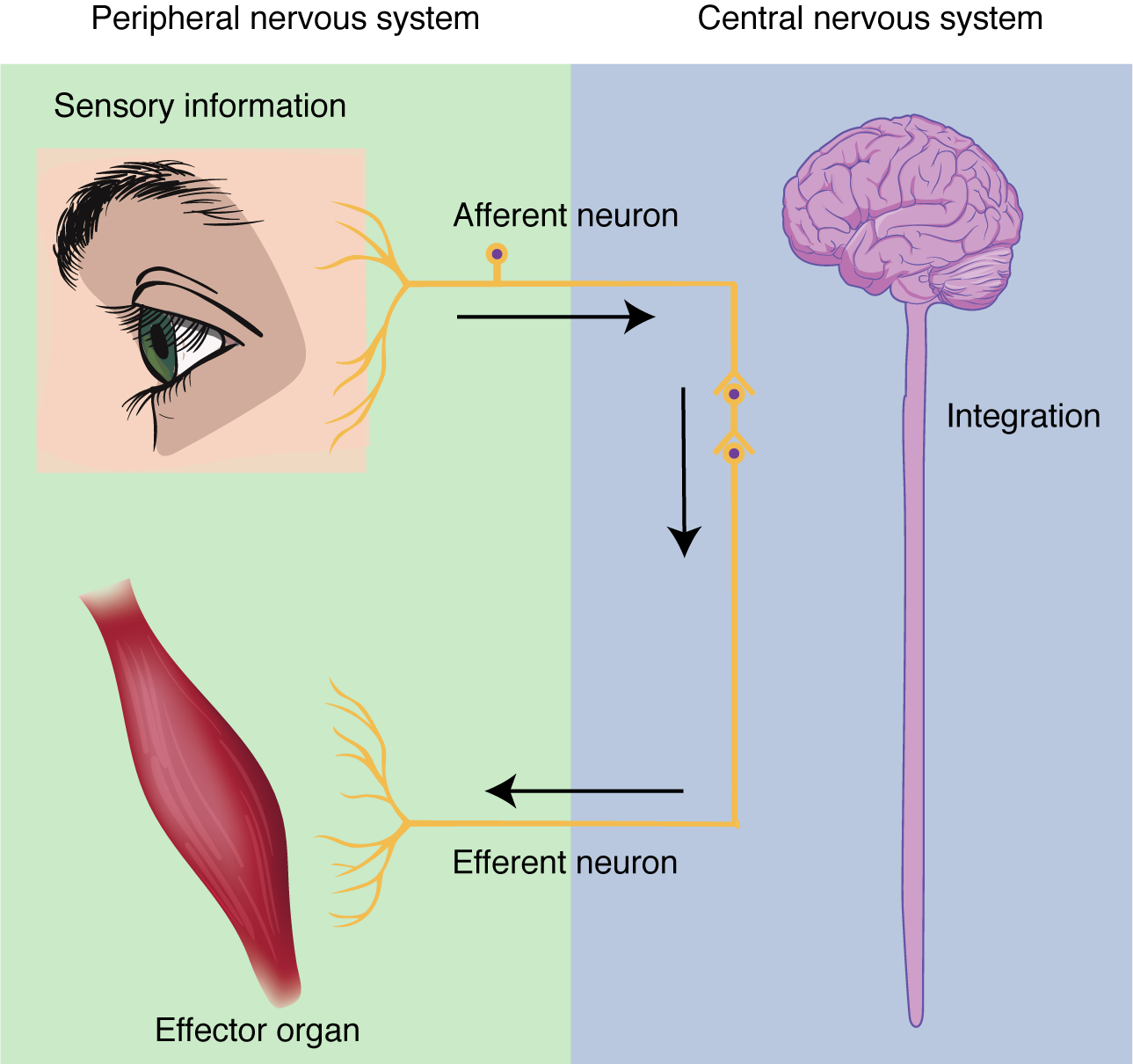
Sensation . Sensation refers to receiving information about the environment, either what is happening outside (ie: heat from the sun) or inside the body (ie: heat from muscle activity). These sensations are known as stimuli (singular = stimulus ) and different sensory receptors are responsible for detecting different stimuli. Sensory information travels towards the CNS through the PNS nerves in the specific division known as the afferent (sensory) branch of the PNS. When information arises from sensory receptors in the skin, skeletal muscles, or joints, it is transmitted to the CNS using somatic sensory neurons; when information arises from sensory receptors in the blood vessels or internal organs, it is transmitted to the CNS using visceral sensory neurons.
Response. The nervous system produces a response in effector organs (such as muscles or glands) due to the sensory stimuli. The motor ( efferent ) branch of the PNS carries signals away from the CNS to the effector organs. When the effector organ is a skeletal muscle, the neuron carrying the information is called a somatic motor neuron; when the effector organ is cardiac or smooth muscle or glandular tissue, the neuron carrying the information is called an autonomic motor neuron. Voluntary responses are governed by somatic motor neurons and involuntary responses are governed by the autonomic motor neurons, which are discussed in the next section.
Integration . Stimuli that are detected by sensory structures are communicated to the nervous system where information is processed. In the CNS, information from some stimuli is compared with, or integrated with, information from other stimuli or memories of previous stimuli. Then, a motor neuron is activated to initiate a response from the effector organ. This process during which sensory information is processed and a motor response generated is called integration (see Figure 12.1.2 below).
Chapter Review
The nervous system can be separated into divisions on the basis of anatomy and physiology. The anatomical divisions are the central and peripheral nervous systems. The CNS is the brain and spinal cord. The PNS is everything else and includes afferent and efferent branches with further subdivisions for somatic, visceral and autonomic function. Functionally, the nervous system can be divided into those regions that are responsible for sensation, those that are responsible for integration, and those that are responsible for generating responses.
Review Questions
Critical thinking questions.
1. What responses are generated by the nervous system when you run on a treadmill? Include an example of each type of tissue that is under nervous system control.
2. When eating food, what anatomical and functional divisions of the nervous system are involved in the perceptual experience?
Answers for Critical Thinking Questions
- Running on a treadmill involves contraction of the skeletal muscles in the legs (efferent somatic motor), increase in contraction of the cardiac muscle of the heart (efferent autonomic motor), and the production and secretion of sweat in the skin to stay cool (sensation of temp = afferent visceral sensory, sweat gland activation = efferent autonomic motor).
- The perceptual experience of eating food refers to tasting food, both in terms of flavors and texture. The neurons responsible for sensing taste are afferent somatic neurons of the PNS.
This work, Anatomy & Physiology, is adapted from Anatomy & Physiology by OpenStax , licensed under CC BY . This edition, with revised content and artwork, is licensed under CC BY-SA except where otherwise noted.
Images, from Anatomy & Physiology by OpenStax , are licensed under CC BY except where otherwise noted.
Access the original for free at https://openstax.org/books/anatomy-and-physiology/pages/1-introduction .
Anatomy & Physiology Copyright © 2019 by Lindsay M. Biga, Staci Bronson, Sierra Dawson, Amy Harwell, Robin Hopkins, Joel Kaufmann, Mike LeMaster, Philip Matern, Katie Morrison-Graham, Kristen Oja, Devon Quick, Jon Runyeon, OSU OERU, and OpenStax is licensed under a Creative Commons Attribution-ShareAlike 4.0 International License , except where otherwise noted.
- History & Society
- Science & Tech
- Biographies
- Animals & Nature
- Geography & Travel
- Arts & Culture
- Games & Quizzes
- On This Day
- One Good Fact
- New Articles
- Lifestyles & Social Issues
- Philosophy & Religion
- Politics, Law & Government
- World History
- Health & Medicine
- Browse Biographies
- Birds, Reptiles & Other Vertebrates
- Bugs, Mollusks & Other Invertebrates
- Environment
- Fossils & Geologic Time
- Entertainment & Pop Culture
- Sports & Recreation
- Visual Arts
- Demystified
- Image Galleries
- Infographics
- Top Questions
- Britannica Kids
- Saving Earth
- Space Next 50
- Student Center
- Introduction
Human nervous system interactive
Neuronal development.
- Morphological development
- Postnatal changes
- Lobes of the cerebral cortex
- Cerebral ventricles
- Basal ganglia
- Epithalamus
- Hypothalamus
- Subthalamus
- Medulla oblongata
- Cellular laminae
- Dorsal column
- Spinothalamic tracts
- Spinocerebellar tracts
- Corticospinal tract
- Rubrospinal tract
- Vestibulospinal tract
- Reticulospinal tract
- Autonomic tracts
- Structural components of spinal nerves
- Functional types of spinal nerves
- Cervical plexus
- Brachial plexus
- Lumbar plexus
- Sacral plexus
- Coccygeal plexus
- Olfactory nerve (CN I or 1)
- Optic nerve (CN II or 2)
- Oculomotor nerve (CN III or 3)
- Trochlear nerve (CN IV or 4)
- Ophthalmic nerve
- Maxillary nerve
- Mandibular nerve
- Abducens nerve (CN VI or 6)
- Facial nerve (CN VII or 7)
- Vestibulocochlear nerve (CN VIII or 8)
- Glossopharyngeal nerve (CN IX or 9)
- Vagus nerve (CN X or 10)
- Accessory nerve (CN XI or 11)
- Hypoglossal nerve (CN XII or 12)
- Sympathetic ganglia
- Neurotransmitters and receptors
- Parasympathetic nervous system
- Enteric nervous system
- Reflex actions
- Tendon organs
- Muscle spindles
- Stretch reflexes
- Reciprocal innervation
- Lower-level mechanisms of movement
- Cerebral hemispheres
- Saccule and utricle
- Semicircular canals
- Nerve supply
- Vestibulo-ocular reflex
- Conscious sensation
- The urinary system
- The reproductive system
- The endocrine system
- Reflex pathways
- Vasopressin and cardiovascular regulation
- Theories of pain
- Peripheral nerves
- Spinal cord
- Central pain
- Referred pain
- Changes in the cerebral cortex
- General organization of perception
- The defense reaction
- Urination and defecation
- Eating and drinking
- Temperature regulation
- Reward and punishment
- Circadian rhythms
- Analytical approaches
- Hemispheric asymmetry, handedness, and cerebral dominance
- Executive functions of the frontal lobes

human nervous system
Our editors will review what you’ve submitted and determine whether to revise the article.
- Table Of Contents

human nervous system , system that conducts stimuli from sensory receptors to the brain and spinal cord and conducts impulses back to other parts of the body. The conduction of electrochemical stimuli from sensory receptors occurs via organized groups of specialized cells, consisting largely of neurons , various neural support cells, and tracts of nerve fibers, which serve as a network channeling neural impulses to the site at which a response occurs.
As with other higher vertebrates, the human nervous system has two main parts: the central nervous system (the brain and spinal cord) and the peripheral nervous system (the nerves that carry impulses to and from the central nervous system). In humans the brain is especially large and well developed.
Prenatal and postnatal development of the human nervous system
Almost all nerve cells, or neurons , are generated during prenatal life, and in most cases they are not replaced by new neurons thereafter. Morphologically, the nervous system first appears about 18 days after conception , with the genesis of a neural plate . Functionally, it appears with the first sign of a reflex activity during the second prenatal month, when stimulation by touch of the upper lip evokes a withdrawal response of the head. Many reflexes of the head, trunk, and extremities can be elicited in the third month.

During its development the nervous system undergoes remarkable changes to attain its complex organization. In order to produce the estimated 1 trillion neurons present in the mature brain, an average of 2.5 million neurons must be generated per minute during the entire prenatal life. This includes the formation of neuronal circuits comprising 100 trillion synapses , as each potential neuron is ultimately connected with either a selected set of other neurons or specific targets, such as sensory endings. Moreover, synaptic connections with other neurons are made at precise locations on the cell membranes of target neurons. The totality of these events is not thought to be the exclusive product of the genetic code , for there are simply not enough genes to account for such complexity. Rather, the differentiation and subsequent development of embryonic cells into mature neurons and glial cells are achieved by two sets of influences: (1) specific subsets of genes and (2) environmental stimuli from within and outside the embryo. Genetic influences are critical to the development of the nervous system in ordered and temporally timed sequences. Cell differentiation, for example, depends on a series of signals that regulate transcription, the process in which deoxyribonucleic acid ( DNA ) molecules give rise to ribonucleic acid ( RNA ) molecules, which in turn express the genetic messages that control cellular activity. Environmental influences derived from the embryo itself include cellular signals that consist of diffusible molecular factors ( see below Neuronal development ). External environmental factors include nutrition, sensory experience, social interaction, and even learning. All of these are essential for the proper differentiation of individual neurons and for fine-tuning the details of synaptic connections. Thus, the nervous system requires continuous stimulation over an entire lifetime in order to sustain functional activity.

In the second week of prenatal life, the rapidly growing blastocyst (the bundle of cells into which a fertilized ovum divides) flattens into what is called the embryonic disk . The embryonic disk soon acquires three layers: the ectoderm (outer layer), mesoderm (middle layer), and endoderm (inner layer). Within the mesoderm grows the notochord , an axial rod that serves as a temporary backbone. Both the mesoderm and notochord release a chemical that instructs and induces adjacent undifferentiated ectoderm cells to thicken along what will become the dorsal midline of the body, forming the neural plate. The neural plate is composed of neural precursor cells, known as neuroepithelial cells, which develop into the neural tube ( see below Morphological development ). Neuroepithelial cells then commence to divide, diversify, and give rise to immature neurons and neuroglia, which in turn migrate from the neural tube to their final location. Each neuron forms dendrites and an axon ; axons elongate and form branches, the terminals of which form synaptic connections with a select set of target neurons or muscle fibers.

The remarkable events of this early development involve an orderly migration of billions of neurons , the growth of their axons (many of which extend widely throughout the brain), and the formation of thousands of synapses between individual axons and their target neurons. The migration and growth of neurons are dependent, at least in part, on chemical and physical influences. The growing tips of axons (called growth cones) apparently recognize and respond to various molecular signals, which guide axons and nerve branches to their appropriate targets and eliminate those that try to synapse with inappropriate targets. Once a synaptic connection has been established, a target cell releases a trophic factor (e.g., nerve growth factor ) that is essential for the survival of the neuron synapsing with it. Physical guidance cues are involved in contact guidance, or the migration of immature neurons along a scaffold of glial fibers.
In some regions of the developing nervous system, synaptic contacts are not initially precise or stable and are followed later by an ordered reorganization, including the elimination of many cells and synapses. The instability of some synaptic connections persists until a so-called critical period is reached, prior to which environmental influences have a significant role in the proper differentiation of neurons and in fine-tuning many synaptic connections. Following the critical period, synaptic connections become stable and are unlikely to be altered by environmental influences. This suggests that certain skills and sensory activities can be influenced during development (including postnatal life), and for some intellectual skills this adaptability presumably persists into adulthood and late life.
Got any suggestions?
We want to hear from you! Send us a message and help improve Slidesgo
Top searches
Trending searches

mexican independence
54 templates

hispanic heritage month
21 templates

49 templates

22 templates

independencia de mexico
14 templates

indigenous canada
47 templates
Learn More About the Nervous System
It seems that you like this template, learn more about the nervous system presentation, free google slides theme, powerpoint template, and canva presentation template.
Discover the fascinating world of the nervous system with this creative template. With calming colors and engaging visuals, you'll be able to explore the different components of the nervous system in a fun and informative way. Plus, you'll get an in-depth look at the neurons, neurotransmitters, and the effects of certain hormones on the nervous system. Take your audience on an exciting journey into the science of the nervous system. Download this design to learn more about the nervous system now!
Features of this template
- 100% editable and easy to modify
- 35 different slides to impress your audience
- Contains easy-to-edit graphics such as graphs, maps, tables, timelines and mockups
- Includes 500+ icons and Flaticon’s extension for customizing your slides
- Designed to be used in Google Slides, Canva, and Microsoft PowerPoint
- 16:9 widescreen format suitable for all types of screens
- Includes information about fonts, colors, and credits of the resources used
How can I use the template?
Am I free to use the templates?
How to attribute?
Combines with:
This template can be combined with this other one to create the perfect presentation:

Attribution required If you are a free user, you must attribute Slidesgo by keeping the slide where the credits appear. How to attribute?

Register for free and start downloading now
Related posts on our blog.

How to Add, Duplicate, Move, Delete or Hide Slides in Google Slides

How to Change Layouts in PowerPoint

How to Change the Slide Size in Google Slides
Related presentations.

Create your presentation Create personalized presentation content
Writing tone, number of slides.

Premium template
Unlock this template and gain unlimited access

Register for free and start editing online
Ch. 7 – The Nervous System
- Overview & Organization of the Nervous System
Functions of the Nervous System
The master controlling & communicating system of the body…
- Sensory input —gathering information
- To monitor changes occurring inside and outside the body
- Changes = stimuli
- Integration
- To process and interpret sensory input and decide if action is needed
- Motor output
- A response to integrated stimuli
- The response activates muscles or glands
- Structural Classification �of the Nervous System
- Central nervous system (CNS) – dorsal body cavity; integrating and command centers; interpret sensory information & give out instructions
Spinal cord
- Peripheral nervous system (PNS) – outside of CNS
- Nerves outside the brain and spinal cord
- Spinal nerves – carry impulses to and from spinal cord
- Cranial nerves – carry impulses to and from brain
- Functional Classification of �the Peripheral Nervous System
- Sensory (afferent) division
- Nerve fibers that carry information to the CNS
- Somatic sensory fibers – deliver impulses from skin, skeletal muscle, and joints
- Visceral sensory fibers (afferents) – deliver impulses from viscera
- Motor (efferent) division
- Nerve fibers that carry impulses away from the CNS
- Somatic (voluntary) NS – voluntary control of skeletal muscles
- Autonomic (involuntary NS – involuntary control of smooth & cardiac muscle and glands
- Divided into sympathetic and parasympathetic NS
Answer Did You Get It? #1
- Structure & Function of Nervous Tissue
- Support Cells
- Support cells in the CNS are grouped together as neuroglia (AKA glia or glial cells ) = “nerve glue”
- Functions: support, insulate, and protect neurons
- Cannot transmit nerve impulses (as can neurons)
- Never lose their ability to divide (as neurons do)
- Most brain tumors are gliomas
- Glia of the Central Nervous System:
- Ependymal cells
- Oligodendrocytes
- Glia of the Peripheral Nervous System:
- Schwann cells
- Satellite cells
Support Cells, continued…
- Abundant (~1/2 of neural tissue)
- Star-shaped cells
- Brace & anchor neurons to capillaries
- Form living barrier between capillaries and neurons (exchange) (blood-brain barrier)
- Control brain’s chemical environment
- Absorb leaked K + ions
- Absorb released neurotransmitters
- Spiderlike phagocytes
- Protect from infection
- Dispose of debris
- Dead brain cells & bacteria
- Line cavities of the brain and spinal cord
- Beating cilia circulate cerebrospinal fluid (CSF)
- CSF fills brain & spinal cord cavities & serves as cushion
- Wrap around nerve fibers in the CNS
- Produce fatty insulating coverings = myelin sheaths
- Protect neuron cell bodies
- Form myelin sheath around nerve fibers in the PNS
Answer Did You Get It? #’s 2-3
- Neurons = nerve cells
- Cells specialized to transmit nerve impulses from one part of body to another
- Two major regions of neurons:
- Metabolic center: contains nucleus, large nucleolus
- No centrioles = no mitosis
- Nissl substance = specialized RER
- Neurofibrils (intermediate cytoskeleton)
- Maintain cell shape
Neurons, continued…
- Processes outside the cell body
- Microscopic to 3-4 ft in length
- Longest = from lumbar region of spine to great toe
- Dendrites —conduct impulses toward the cell body
- A neuron may have hundreds
- Axons —conduct impulses away from the cell body
- Arises from cone-like region of cell body called axon hillock
- Collateral branches
- End in highly branched axon terminals
- Axon terminals contain vesicles with neurotransmitters
- Axonal terminals are separated from the next neuron by a synaptic cleft
- Synapse —junction between nerves ( syn = clasp/join)
Neuron processes, continued…
- Myelin sheath —whitish, fatty material covering axons
- Protects & insulates fibers
- Increases rate of nerve impulse transmission
- Schwann cells —produce myelin sheaths in jelly roll–like fashion
- Schwann cells in the PNS; oligodendrocytes in the CNS
- Neurilemma – portion of cell membrane on outer layer of coil where most of its cytoplasm resides
- Nodes of Ranvier —gaps in myelin sheath along the axon
- Aid in speeding up nerve impulses – saltatory conduction
- Homeostatic imbalance – multiple sclerosis = gradual destruction of myelin sheaths (become hardened = sclerosis), autoimmune disease (sheath protein)
- Visual & speech disturbances, loss of muscle control, increasingly disabled
- Interferon injections provide relief; no cure
- Terminology of Neurons
- Most neuron cell bodies are found in the CNS
- Nuclei —clusters of cell bodies within the white matter of the CNS (protected within the brain case and vertebral column)
- Ganglia —small collections of cell bodies in the PNS
- Tracts = bundles of nerve fibers in CNS
- White matter – myelinated tracts in CNS
- Gray matter —cell bodies and unmyelinated tracts in CNS
- Nerves = bundles of nerve fibers in PNS
- Functional Classification of Neurons
Direction of nerve impulse with respect to CNS
- Sensory (afferent) neurons
- Carry impulses from the sensory receptors to the CNS
- Ganglion outside of CNS
- Dendrite endings associate with receptors
- Cutaneous sense organs in muscles and tendons
- Proprioceptors —detect stretch or tension
Naked nerve ending; pain/temp
Meissner’s corpuscule: touch
Pacinian corpuscule: deep pressure
Golgi tendon organ & muscle spindle;: proprioception
Functional Classification of Neurons, continued…
- Motor (efferent) neurons
- Carry impulses from the central nervous system to viscera, muscles, or glands
- Cell bodies always in CNS
- Interneurons (association neurons)
- Connect sensory and motor neurons in neural pathways
- Structural Classification of Neurons
- Multipolar neurons—many extensions from the cell body
- most common
- Bipolar neurons—one axon and one dendrite
- Rare in adults
- Act in sensory processing – eye, nose
- Unipolar neurons—have a short single process leaving the cell body
- Divides into proximal (central) and distal (peripheral) processes
- Dendrites only at peripheral end
- Conducts action potentials both ways
- Found in sensory neurons of PNS ganglia
Answer Did You Get It? #’s 4-7
- Physiology of the Nervous System
- Functional Properties of Neurons
- Irritability - ability to respond to stimuli and convert to nerve impulses
- Conductivity - ability to transmit an impulse to other neurons, muscles, or glands
- Nerve Impulses
- Electrical conditions of a resting neuron’s membrane
- Polarized – resting/inactive neuron
- Fewer positive ions on inner face of plasma membrane than on outer face
- Depolarized – stimulated neuron
- More positive ions inside the cell than outside
Nerve Impulses, continued…
- Action Potential Initiation and Generation
- Stimuli excite neurons: light, sound, pressure, mostly neurotransmitters released by other neurons
- Cause a temporary change in the cell membrane’s permeability
- Stimulus causes sodium channel gates to open, and sodium to rush in
- Causes depolarization of the neuron’s membrane
- Inside more positive, outside less positive = graded/local potential
- If stimulus is strong enough, a long distance signal called an action potential or nerve impulse occurs
- Nerve impulses are all-or-nothing responses – they are either propagated over the entire axon or not at all
- Repolarization
- Membrane immediately becomes impermeable to sodium, but permeable to potassium ions
- K + ions rush out of the neuron, restoring electrical conditions to polarized = repolarization
- Repolarization must occur before another impulse can be conducted
- The sodium-potassium pump, using ATP, restores the original concentrations of Na + and K + .
- Saltatory conduction = In myelinated fibers, propagation occurs more quickly since the nerve impulse jumps from node to node.
- Homeostatic imbalance: factors that impair impulse conduction:
- Sedatives & anesthetics block sodium entry
- Cold & continuous pressure interrupt blood circulation (nutrients & O 2 ) – e.g. ice creates numbness, foot “goes to sleep”; prickly feeling caused by impulse transmission starting back up
- Transmission of the Signal at Synapses
- Neurotransmitter is released from vesicles within the axon terminal
- Neurotransmitter molecules diffuse across the synapse
- Neurotransmitters bind to receptors in the membrane of the next neuron
- If enough neurotransmitters are released, another nerve impulse will be generated in this neuron
- Enzymes remove the neurotransmitters from the receptors
- Impulse transmission is an electrochemical event – electrical along the neuron’s membrane; chemical within the synapses
Axon�terminal
Synaptic�cleft
Action�potential�arrives
Axon of�transmitting�neuron
Receiving�neuron
Neurotrans-�mitter is re-�leased into�synaptic cleft
Neurotrans-�mitter binds�to receptor�on receiving�neuron’s�membrane
Vesicle�fuses with�plasma�membrane
Synaptic cleft
Neurotransmitter�molecules
Ion channels
Receiving neuron
Transmitting neuron
Neurotransmitter
Neurotransmitter�broken down�and released
Ion channel opens
Ion channel closes
- Reflex — rapid, predictable, and involuntary response to a stimulus
- Always travel in one direction
- Occurs over pathways called reflex arcs
- Reflex arc — direct route from a sensory neuron, to an interneuron, to an effector
- Neural pathway involving the CNS and PNS
Stimulus at distal�end of neuron
(in cross section)
Interneuron
Sensory neuron
Motor neuron
Integration�center
Reflexes, continued…
- Types of Reflexes
- Somatic reflexes
- Reflexes which stimulate the skeletal muscles
- Example: moving hand away from a hot stove
- Autonomic reflexes
- Regulate the activity of smooth muscles, heart, and glands
- Examples: salivary reflex, pupillary reflex
- Regulate: digestion, elimination, blood pressure, and sweating
- Parts of a reflex arc
- Sensory receptor – reacts to a stimulus
- Integration center
- Effector organ – muscle or gland which is stimulated
- Patellar (knee-jerk) reflex is an example of a two-neuron reflex arc
Figure 7.11d
Figure 7.11b–c
Sensory (afferent)�neuron
Motor�(efferent)�neuron
Sensory receptors�(stretch receptors�in the quadriceps�muscle)
Effector�(quadriceps�muscle of�thigh)
Synapse in�ventral horn�gray matter
Inter-�neuron
Sensory receptors�(pain receptors in�the skin)
Effector�(biceps�brachii�muscle)
- Flexor (withdrawal) reflex is an example of a three-neuron reflex arc
- Withdrawal reflex arc has an interneuron
- The more neurons involved, the slower the communication because of the time it takes for neurotransmitters to diffuse
- Many spinal reflexes do not involve the brain
- Other reflexes require the brain to evaluate different types of information
- Reflex testing evaluates condition of the nervous system
- Exaggerated, distorted, and absent reflexes indicate nervous system disorders
Answer Did You Get It? #’s 8-11
- Central Nervous System (CNS)
- CNS develops from the embryonic neural tube
- Runs along the dorsal median plane
- 4 th week – anterior end expands = brain formation
- Rest of tube = spinal cord
- The central canal of the neural tube enlarges into 4 chambers = ventricles
- Filled with cerebrospinal fluid
- Functional Anatomy of the Brain
- ~3 lbs, wrinkled, texture similar to cold oatmeal
- 4 major regions:
- Cerebral hemispheres (cerebrum)
- Diencephalon
Regions of the Brain: Cerebrum
- Cerebrum (cerebral hemispheres)
- Paired, superior parts of the brain
- Includes more than half of the brain mass; obscures most of the brain stem
- The surface is made of ridges ( gyri = “twisters”) and grooves ( sulci = “furrows”)
- Fissures (deep grooves) divide the cerebrum into lobes
- Occipital lobe
- Temporal lobe
Figure 7.13b
- Cerebral Cortex
- Functions : speech, memory, logic, emotion, consciousness, sensation interpretation, & voluntary movement
- Cell bodies of neurons in cerebral cortex in outermost gray matter
- Primary somatic sensory area
- In parietal lobe posterior to central sulcus
- Receives & interprets impulses from the body’s sensory receptors
- Detects: pain, cold, light touch
Sensory & motor homunculus – the more neurons there are for a function, the larger the area represented by that body region
Figure 7.14
- Visual area in occipital lobe
- Auditory area in temporal lobe
- Olfactory area deep in temporal lobe
- Primary motor area in frontal lobe
- Conscious movement of skeletal muscle
- Axons of these motor neurons form the corticospinal or pyramidal tract
- Descends to spinal cord
- Broca’s area at base of precentral gyrus
- Involved in our ability to speak
- Only located in one (usually left) hemisphere
- Damage here can cause inability to speak – conscious of what you want to say, but unable to do it
- Frontal association areas – higher intellectual reasoning & socially acceptable behavior
- Complex memories stored in temporal and frontal lobes
- Speech/language (Wernicke’s) area – junction of temporal, parietal, & occipital lobes
- Allows us to sound out words
- Usually in just one hemisphere
- Damage: Wernicke’s aphasia – lack of language comprehension; clear speaking though
- Frontal lobes – language comprehension (word meaning)
- Gustatory area – taste – base of primary somatic sensory area (parietal)
- General interpretation area – temporal & parietal
- Cerebral White Matter
- White matter—fiber tracts carrying impulses to, from, and within the cortex
- Corpus callosum – large tract connecting hemispheres; allows hemispheres to communicate with one another
- Called commisures
- Association fiber tracts connect areas within hemispheres ; projection fiber tracts connect cerebrum to lower CNS centers
- Basal nuclei (basal ganglia ) — islands of gray matter buried within the white matter
- Regulate voluntary
motor activities
- Homeostatic Imbalance:
- Problems with basal
nuclei cause difficulty in
walking or other voluntary
movements: Huntington’s
disease & Parkinson’s
Answer Did You Get It? #’s 12-13
- Regions of the Brain: Diencephalon (Interbrain)
- Sits on top of brain stem; enclosed by the cerebral hemispheres
- Made of three parts: Thalamus, Hypothalamus, Epithalamus
- Thalamus – relay station for sensory impulses traveling up to sensory cortex
- Crude awareness of a pending sensation being pleasant or not
- Hypothalamus – floor of diencephalon
- Autonomic NS center: helps body temp, water balance, & metabolism
- Limbic system – “emotional-visceral brain” where thirst, appetite, sex, pain, and pleasure centers are
- Regulates the pituitary gland ; secretes hormones
- Mammillary bodies – reflex centers involved in olfaction
Regions of the Brain: Diencephalon
- Epithalamus
- Forms the roof of the third ventricle
- Houses the pineal body (an endocrine gland)
- Includes the choroid plexus —complex of capillaries which form cerebrospinal fluid
Regions of the Brain: Brain Stem
- Small: ~thumb in diameter & ~3” long
- 3 regions: midbrain, pons, & medulla oblongata
- Provides a pathway for ascending & descending tracts
- Contains nuclei with rigidly programmed autonomic behaviors necessary for survival
- Some connected to cranial nerves controlling breathing & blood pressure
- From mammilary bodies to pons
- Cerebral aqueduct – canal connecting 3 rd ventricle of diencephalon to 4 th ventricle
- Has two bulging fiber tracts — cerebral peduncles : convey ascending & descending impulses
- Mostly composed of tracts of nerve fibers
- Has four rounded protrusions— corpora quadrigemina (“gemini” = twins)
- Reflex centers for vision and hearing
- Pons (“bridge”)
- Rounded part of brain stem just below midbrain
- Mostly composed of fiber tracts
- Includes nuclei involved in the control of breathing
- Medulla Oblongata
- Most inferior part of the brain stem
- Merges into the spinal cord
- Includes important fiber tracts
- Contains nuclei which control:
- Blood pressure
- Fourth ventricle
- Reticular Formation
- Diffuse mass of gray matter along the length of the brain stem
- Involved in motor control of visceral organs
- Reticular activating system (RAS) plays a role in awake/sleep cycles and consciousness
- Damage here can cause a coma (permanent unconsciousness)
- Regions of the Brain: Cerebellum
- Cauliflower-like, dorsally projecting from under the occipital lobe
- Two hemispheres with convoluted surfaces
- Outer cortex composed of gray matter; inner region composed of white matter
- Provides precise timing for skeletal muscle activity and controls balance & equilibrium
- “Automatic pilot” – compares brain’s intentions with body’s actual performance; initiates appropriate corrective measures
- Ataxia – damage to cerebellum can result in clumsy & disorganized movements; appear to be drunk
Answer Did You Get It? #’s 14-16
- Protection of the Central Nervous System
- Nervous tissue is soft and delicate; neurons injured easily
- Brain and spinal cord protected by
- Scalp and skin
- Skull and vertebral column
- Meninges (membranes)
- Cerebrospinal fluid (watery cushion)
- Blood-brain barrier – protection from harmful substances in the blood
Figure 7.17b
- Connective tissue membranes which cover & protect the CNS
- Double-layered, outermost layer; leathery
- Periosteal layer (periosteum)—attached to inner surface of the skull
- Meningeal layer —outer covering of the brain; fuses with the dura mater of the spinal cord
- Layers are fused except in dural venous sinuses where venous blood is collected
- Inward folds attach brain to cranial cavity
- Falx cerebri & tantorium cerebelli
- Arachnoid mater (“spider”)
- Middle layer
- Attached to the pia mater, forming the subarachnoid space
- Filled with cerebrospinal fluid (CSF)
- Arachnoid villi – projections of arachnoid mater; protrude through dura mater
- CSF passes into dural sinuses through these structures
- Pia mater (“gentle mother”)
- Innermost membrane
- Clings tightly brain and spinal cord surfaces
- Epidural injections – “upon the dura”
- Homeostatic Imbalance :
- Meningitis – inflammation of the meninges
- Bacterial or vial infections
- Serious threat to brain if spreads into CNS
- Encephalitis – inflammation of the brain
- Diagnosed by sampling CSF
Cerebrospinal Fluid (CSF)
- Similar to blood plasma composition
- Less protein, more vitamin C, different ion composition
- Formed from blood by choroid plexuses
- Clusters of capillaries hanging from each of brain’s ventricles
- Forms a watery cushion to protect the brain from trauma
- Circulated in arachnoid space, ventricles, and central canal of the spinal cord
- CSF continually circulates in brain
- From two lateral ventricles, to 3 rd ventricle, through cerebral aqueduct, to 4 th ventricle
- Some CSF continues to spinal cord
- Normally circulates at a constant rate
- Changes to CSF composition may indicate meningitis, tumors, or MS
- Lumbar/spinal tap – sample the CSF
- Remain lying down for 12 hrs or “spinal headache”
- Homeostatic Imbalance - Hydrocephalus
- If something obstructs CSF drainage, it accumulates and exerts pressure on the brain
- “Water on the brain”
- Results in enlarged head in newborns with increasing brain size
- Would cause brain damage in adults
- Treated by surgically inserting a shunt (plastic drain); drains excess fluid into a vein
- Blood-Brain Barrier
- Brain is super sensitive to having a constant internal environment
- Neurons kept separated from bloodborne substances by the blood-brain barrier
- Composed of least permeable capillaries of the body
- Bound by tight junctions
- Allowed to enter:
- Water, glucose, and essential amino acids pass easily through
- Metabolic wastes (urea, toxins, proteins, most drugs), nonessential amino acids, K +
- Useless as a barrier against some substances
- Fats and fat soluble molecules
- Respiratory gases
Answer Did You Get It? #’s 17-19
- Traumatic Brain Injuries
- Head injuries are leading cause of accidental death in US; caused by damaging blow to head
- Further damage caused by brain ricocheting on opposite end of skull
- Slight brain injury
- Dizzy/”see stars,” briefly lose consciousness
- No permanent brain damage
- Marked tissue destruction occurs
- May remain conscious if cerebral cortex injury; may be in coma if brain stem is injured severely (especially RAS)
- Nervous tissue does not regenerate
- Intracranial hemorrhage
- Bleeding from ruptured vessels
- May cause death
- Cerebral edema
- Brain swelling from the inflammatory response
- May compress and kill brain tissue – neurological deterioration
- Cerebrovascular Accident (CVA/Stroke)
- 3 rd leading cause of death in US
- Blood circulation to brain is obstructed by a blood clot or ruptured blood vessel
- Brain tissue supplied with oxygen from that blood source dies
- Loss of some functions or death may result; undamaged neurons can spread into damaged areas and take over some lost functions (= neuroplasticity )
- Hemiplegia – one-sided paralysis ( e.g. right-sided paralysis = damage to left motor cortex)
- Apahsia – damage to language areas
- Motor/Broca’s aphasia – loss of ability to speak
- Sensory/Wernicke’s aphasia – loss of ability to understand written & spoken language
- Transient ischemic attack (ITA) – temporary restriction of blood flow (ischemia) to brain
- Last 5-50 min; numbness, temporary paralysis; impaired speech
- Warning of impending, more serious CVA
Answer Did You Get It #20
- The Terrible Three
- Alzheimer’s Disease
- Progressive degenerative brain disease, results in dementia (mental deterioration)
- Mostly seen in the elderly, but may begin in middle age
- Victims experience: memory loss, short attention span, disorientation, eventual loss of language, irritability, moodiness, confusion, sometimes violent, and ultimately, hallucinations.
- Structural changes in the brain include: low Ach, shrinking gyri, brain atrophy (especially in areas of thought and memory), abnormal protein (senile plaque – beta amyloid peptide ) deposits, and twisted tau fibers within neurons
- Treat with acetylcholinesterase inhibitors
- Parkinson’s Disease
- Problem associated with basal nuclei; cause not known
- Typically affects people in 50’s-60’s
- Degeneration of dopamine-releasing neurons in the substantia nigra, causing basal nuclei to become overactive
- Symptoms: persistent tremor (even at rest), head nodding, “pill-rolling” of fingers, forward-bent walking posture, shuffling gait, stiff facial expressions, difficulty in initiating movements
- Treatments: L-dopa for some symptoms (bad side effects); deprenyl to slow degeneration; thalamic stimulation via electrodes alleviates tremors; implants of embryonic tissue promising
- Huntington’s Disease
- Genetic disorder (dominant) – typically occurs at middle-age
- Massive degeneration of basal nuclei and later of the cerebral cortex
- Progressive symptoms: wild, jerky movements ( chorea ), later marked mental deterioration
- Typically fatal within 15 years
- Overstimulation of motor cortex
- Treat with drugs that block dopamine; fetal tissue implants are promising
- Spinal Cord
- 2-way conduction pathway to and from the brain
- Major reflex center (spinal reflexes)
- Extends from the foramen magnum of the skull to the first or second lumbar vertebra
- Cushioned & protected by meninges
- 31 pairs of spinal nerves arise from the spinal cord
- Cervical & lumbar enlargements – origin of upper & lower limb nerves
- Cauda equina (horse’s tail) is a collection of spinal nerves at the inferior end
Spinal Cord Anatomy
- Gray matter of Spinal Cord and Spinal Roots
- Gray matter surrounds the central canal (filled with CSF)
- Dorsal (posterior) horns – project posteriorly
- Contain interneurons
- Sensory neuron cell bodies in dorsal root ganglia ; enter spinal cord through dorsal root
- Anterior (ventral) horns – project anteriorly
- Motor neuron cell bodies in ventral horns; axons exit spinal cord through ventral root
- Homoeostatic imbalance – flaccid paralysis – damage to ventral root = no stimulation of muscles
- Spinal nerves – fusion of dorsal and ventral roots
- White matter of the Spinal Cord
- Myelinated fiber tracts (see 7.22)
- Dorsal, lateral, ventral columns
- Sensory/afferent tracts – conduct sensory impulses to brain
- Motor/efferent tracts – conduct impulses from brain to skeletal muscles
- Dorsal column tracts are all ascending carrying sensory input to brain
- Lateral & ventral tracts contain both ascending & descending tracts
- Homeostatic imbalance – spastic paralysis : transected (cut crosswise) or crushed spinal cord – affected muscles stay healthy b/c still stimulated, but moments become spastic; loss of feeling below injury
- Quadriplegic = 4 limbs affected
- Paraplegic = legs only
Answer Did You Get It? #’s 21-23
- Peripheral Nervous System (PNS)
- Nerves and ganglia outside CNS
- Structure of a Nerve
- Nerve = bundle of neuron fibers outside the CNS
- Neuron fibers are bundled by connective tissue
- Delicate endoneurium surrounds each fiber
- Groups of fibers are bound into fascicles by coarser perineurium
- Fascicles are bound together by tough, fibrous epineurium
- Forms cordlike nerve
Structure of a Nerve, continued…
- Nerves are classified according to the direction in which they transmit impulses:
- Mixed nerves – nerves with both sensory and motor fibers
- Sensory (afferent) nerves – nerves carrying impulses toward the CNS
- Motor (efferent) nerves – nerves carrying impulses away from the CNS
- Cranial Nerves
- 12 pairs of nerves that mostly serve the head and neck
- Only the pair of vagus nerves extend to thoracic and abdominal cavities
- Numbered in order; names typically match the structures they control
- Most are mixed nerves, but three are sensory only (optic, olfactory, & vestibulocochlear)
Cranial Nerves, continued…
- Olfactory nerve — sensory for smell
- Optic nerve — sensory for vision
- Oculomotor nerve — motor fibers to eye muscles (most movements, lens shape, & pupil size)
- Trochlear nerve — motor fiber to eye muscle (superior oblique)
- Trigeminal nerve — sensory for the face, nose, & mouth; motor fibers to chewing muscles
- Abducens nerve — motor fibers to eye muscles (lateral movement)
- Facial nerve — sensory for anterior taste buds; motor fibers for facial expression and lacrimal & salivary glands
- Vestibulocochlear nerve — sensory for balance and hearing
- Glossopharyngeal nerve — sensory for posterior taste buds; motor fibers to the pharynx (swallowing & saliva production); carotid artery pressure sensors
- Vagus nerves — sensory and motor fibers for pharynx, larynx, and thoracic & abdominal viscera (mostly parasympathetic = promote digestion & regulate heart activity)
- Accessory nerve — motor fibers to sternocleidomastoid & trapezius
- Hypoglossal nerve — motor fibers for tongue movements; sensory impulses from tongue
- O h O nce O ne T akes T he A natomy F inal V ery G ood V acations A re H eavenly.
- O nly O wls O bserve T hem T raveling A nd F inding V oldemort G uarding V ery S ecret H orcruxes
- Spinal Nerves & Nerve Plexuses
- There are 31 pairs formed by the combination of the ventral and dorsal roots of the spinal cord
- Named for the region from which they arise
- Spinal nerves divide after leaving the spinal cord
- Dorsal rami — serve the skin and muscles of the posterior trunk
- Ventral rami — for nerves T 1 -T 12 forms intercostal nerves (muscles between ribs & skin and muscles of anterior trunk); for rest of nerves forms a nerve networks ( plexus ) for limb sensory & motor
Answer Did You Get It? #’s 24-27
Spinal Nerves & Nerve Plexuses, continued…
- Cervical plexus – from C 1 –C 5 ventral rami
- Phrenic nerve – diaphragm; shoulder/neck muscles
- Brachial plexus – from C 5 –C 8 and T 1 ventral rami
- Axillary nerve – deltoid muscle, shoulder skin; superior thorax muscles & skin
- Radial nerve – triceps & extensor muscles; upper limb posterior skin
- Median nerve – flexor muscles; forearm skin; some hand muscles
- Musculocutaneous nerve – arm flexor muscles; lateral forearm skin
- Ulnar nerve – some forearm flexor muscles; wrist & hand muscles; hand skin
- Lumbar plexus – from L 1 –L 4 ventral rami
- Femoral nerve – lower abdomen , hip flexors & knee extensors; leg & thigh anteromedial skin
- Obturator nerve – adductor & small hip muscles; medial thigh & hip joint skin
- Sacral plexus – from L 4 –L 5 and S 1 –S 4 ventral rami
- Sciatic nerve – largest nerve in body; splits into two nerves; lower trunk & posterior thigh surface (hip extensors & knee flexors)
- Common fibular nerve – lateral leg & foot
- Tibial nerve – posterior leg & foot
- Superior & inferior gluteal nerves – gluteal muscles
Distribution of Major Peripheral Nerves of the �Upper and Lower Limbs
Spinal Nerve Plexuses
Autonomic Nervous System (AKA Involuntary NS)
- Motor subdivision of the PNS
- Controls body activities automatically
- Special neurons that regulate cardiac muscle, smooth muscle (visceral organs & blood vessels), and glands
- Helps to maintain homeostasis – constantly makes adjustments to keep internal conditions stable
- Consists only of motor nerves
Note the differences between ANS & SNS
Autonomic Nervous System, continued…
- Somatic vs. Autonomic nervous systems (both PNS)
- Different effector organs and neurotransmitters
- Somatic NS has cell bodies in CNS and an axon that extends to the effector organ
- Autonomic NS has a chain of two motor neurons
- Preganglionic axon – 1 st neuron; in the CNS (“before the ganglion”)
- Postganglionic axon – 2 nd neuron; outside of CNS; goes to organ
- Two divisions of ANS
- Sympathetic & parasympathetic division
- Regulate the same organs, but with opposite effects (counterbalance one another)
- Sympathetic division – mobilizes body during extreme situations (“fight vs. flight”)
- Parasympathetic division – rest and digest; unwind & conserve
Brain & Spinal Cord Cranial & Spinal Nerves
Sensory Division Motor Division
(Periphery → CNS) (CNS → Periphery)
Afferent/Incoming Efferent/Outgoing
Cranial Spinal Somatic Motor NS Autonomic NS
Nerves Nerves Voluntary Involuntary
Sympathetic Parasympathetic Enteric
Stimulatory Inhibitory GI
- Anatomy of the Parasympathetic Division
- Originates from brain nuclei of cranial nerves (III, VII, IX, & X) and S 2 -S 4
- AKA craniosacral division
- Cranial neurons synapse with ganglionic motor neuron in terminal ganglia (basically are at the effector organs)
- Sacral preganglionic neurons form pelvic splanchnic nerves (pelvic nerves) – pelvic cavity
- Always uses acetylcholine as a neurotransmitter
- Anatomy of the Sympathetic Division
- Originates from gray matter in spinal cord from T 1 through L 2
- AKA thoracolumbar division
- Ganglia are at the sympathetic trunk (near the spinal cord)
- Short pre-ganglionic neuron and long post-ganglionic neuron transmit impulse from CNS to the effector
- Norepinephrine and epinephrine are neurotransmitters to the effector organs
- Sympathetic Functioning —“fight or flight”
- Response to unusual stimulus
- Takes over to increase activities
- Remember as the “E” division
- Exercise, excitement, emergency, and embarrassment
- Homeostatic Imbalance – excessive sympathetic NS stimulation
- Type A personality – never slows down; may be susceptible to heart disease, high blood pressure, ulcers
- Parasympathetic Functioning —“housekeeping” activites
- Conserves energy (rest & digest)
- Maintains daily necessary body functions
- Remember as the “D” division
- digestion, defecation, and diuresis
Answer Did You Get It? #’s 28-30
- Tracking Down CNS Problems
- EEG – electroencephalography
- Recording of brain neuron’s electrical impulse transmission
- Attach electrodes on scalp
- Record speed of brain waves (unique to each individual)
- Alpha = awake, relaxed state
- Beta = awake, alert state
- Theta = common in children, not normal adults
- Delta = deep sleep
Tracking Down CNS Problems, continued…
- CT, MRI & PET scans
- CT (computed axial tomography) & MRI (magnetic resonance imaging) – easily identify tumors, intracranial lesions, MS plaques & areas of dead brain tissue (infarcts)
- PET scans – localize lesions that cause epileptic sezures; used for Alzheimer’s diagnosis, and in cancer tumor activity
CT Scan: normal vs. tumor
PET Scan: normal vs. Alzheimer’s disease
- Cerebral angiography
- Used to visualize arteries in brain
- Used to guide a catheter carrying clot-busting drugs (tPA)
Cerebral angiogram showing an aneurism
87-year-old man with acute onset left hemiplegia. . The image on the left (A) obtained preoperatively. The image on the right (B) was obtained after intra-arterial thrombolysis.
- Development Aspects of the Nervous System
- The nervous system is formed during the first month of embryonic development; therefore, any maternal infection can have extremely harmful effects
- Maternal measles (rubella) = deafness
- Lack of O 2 for minutes can cause neuron death
- Smoking decreases amount of O 2 in blood; less O 2 to developing fetus’s brain (potentially brain damage)
- Radiation & drugs (alcohol, opiates, cocaine, etc.) can all damage fetal nervous system development
- Homeostatic imbalances :
- Cerebral palsy – poor control and spastic movements of voluntary muscles, seizures, mental retardation, impaired hearing & vision
- Can be caused by lack of O 2 during difficult delivery
- Anencephaly – failure of the cerebrum to develop; cannot hear, see, or process sensory inputs
- Spina bifida – “forked spine”; vertebra fail to completely form; can result in varying degrees of paralysis & loss of bowel and bladder control
Development Aspects of the Nervous System, cont’d
- The hypothalamus is one of the last areas of the brain to develop (regulates body temperature_
- Premature babies can’t thermoregulate well
- Continued growth & maturation of nervous system through childhood
- Myelination: cranial to caudal; proximal to distal
- Brain is maximum weight as young adult
- Neurons then continue to get damaged and die
- Steady decline of brain weight and volume
- Can still learn throughout life; unlimited neural pathways available
- Sympathetic NS becomes less efficient (especially in constricting blood vessels)
- Orthostatic hypotension – pooling of blood in the feet due to lack of activation of vasoconstrictor fibers and lightheadness; common in elderly when they stand up quickly
- Arteriosclerosis (plaque build up in arteries) and high blood pressure result in less O 2 supply to brain
- Can causes senility – forgetfulness, irritability, confusion, and difficulty in concentrating and thinking clearly
- Some drugs, low blood pressure, constipation, poor nutrition, depression, dehydration, and hormone imbalances can cause “reversible senility”
- Professional boxers (& other high impact sports) and chronic alcoholics hasten the effects of aging on the brain
- “Punch drunk” – slurred speech, tremors, abnormal gait, dementia in retired boxers
- Reduced brain size in both
Answer Did You Get It? #’s 31-32

Organization of The Nervous System
Oct 08, 2011
480 likes | 1.12k Views
Organization of The Nervous System. DR. SANAA ALSHAARAWY & PROF. SAEED ABUEL MAKAREM. Objectives. At the end of the lecture, the students should be able to: List the parts of the nervous system. List the function of the nervous system.
Share Presentation
- nervous system
- cerebrospinal fluid
- autonomic nervous system
- peripheral nervous system involves

Presentation Transcript
Organization of The Nervous System DR. SANAA ALSHAARAWY & PROF. SAEED ABUEL MAKAREM
Objectives At the end of the lecture, the students should be able to: • List the parts of the nervous system. • List the function of the nervous system. • Describe the Structural & Functional Organizations. • Define the terms: Nervous tissue, grey matter, white matter, nucleus, ganglion, tract, nerve. • List the parts of the brain. • List the structures protecting the central nervous system.
INTRODUCTION How does the nervous system work ? • The nervous system has three functions: • Collection of sensory input: • Identifies changes occurring inside or outside the body by using sensory receptors. These changes are called stimuli. • Integration: • Processes, analyzes , and interprets these changes and makes decisions. • Motor output, or response by activating muscles or glands (effectors).
CLASSIFICATION I- Anatomical or Structural classification: 1- Central NS • 2- Peripheral NS II- Physiological or Functional classification: • 1-Sensory division (Afferent) • 2-Motor division (Efferent) • Autonomic • Somatic
Structural Organization Two subdivisions: • Central Nervous System (CNS) • Consists of Brain & Spinal cord • Occupies the dorsal body cavity • Acts as the integrating and command centers. • Peripheral Nervous System (PNS) • Consists of nerves, ganglia, receptors. • It is the part of the nervous system outside the CNS.
Functional Organization • Two subdivisions: • Sensory or afferent division: Consists of nerve fibers that convey impulses from receptors located in various parts of the body, to the CNS. • Motor or efferent division: Consists of nerve fibers that convey impulses from the CNS to the effector organs, muscles and glands. • Both sensory and motor subdivisions are further divided into: • Somaticdivision: concerned with skin, skeletal muscles and joints. • Autonomicdivision: concerned with the visceral organs.
The Nervous System It is the major controlling, regulatory & communicating system in the body. It is the center of all mental activityincluding: Thought, Learning, Behavior and Memory. Together with the endocrine system, the nervous system is responsible for regulating and maintaininghomeostasis.
Nervous Tissue • Nervous system is composed of nervous tissue, which contains two types of cells: 1- Nerve cells or neurons 2- Supporting cells or neuroglia (glia). • Nervous system contains millions of neurons that vary in their shape, size, and number of processes. The junction site of two neurons is called a “synapse or relay”. In the synapses the membranes of adjacent cells are in close apposition(contiguity=contact, not continuity).
Neurons It is the basic structural (anatomical), functional and embryological unit of the nervous system. The human nervous system is estimated to contain about 1010. What is neurone? Prof. Saeed Makarem
Ganglion= A group of neurons outside the CNS Nucleus= A group of neurons within the CNS Remember… Tract =A group of nerve fibers (axons) within the CNS Nerve =A group of nerve fibers (axons) outside the CNS
Grey matter, Which contains 1- Cell bodies & 2- Processes of the neurons, 3- Neuroglia and 4- Blood vessels. White matter, Which contains: 1- Processes of the neurons 2- Neuroglia and 3- Blood vessels NO cell bodies in the white matter. Nervous tissue is organized as:
Neuroglia or glia or glial cells • Neuroglia, or gliacells constitute the other major cellular component of the nervous tissue. • It is a specialized connective tissue supporting framework for the nervous system. • Unlike neurones, neuroglia do not have a direct role in information processing but they are essential for the normal functioning of the neurons, they act as supporting and nutrition for neurons.
Most of the processes of the cell body are short with variable numbers and are receptive in function. They are known as Dendrites.
One of these processes leaving the cell body is called the axonwhich carries information away from the cell body. • Axons are highly variable in length and may divide into several branches or collaterals through which information can be distributed to a number of different destinations. • At the end of the axon, specializations called terminal buttonsoccur. • Here information is transferred to the dendrites of other neurones.
Peripheral NS • Spinal nerves supplying the upper or lower limbs form plexuses e.g. brachialor lumbar plexus. • Nerve cell bodies that are aggregated outside the CNS are called GANGLIA
Autonomic Nervous System • Neurones that detect changes and control the activity of the viscera are collectively referred to as the autonomic nervous system. • Its components are present in both the central and peripheral nervous systems.
SYMPATHETIC & PARASYMPATHETIC SYSTEMS • The autonomic nervous system is divided into two anatomically and functionally distinct parts: • Sympathetic:Or • Thoracolumbar outflow • Parasympathetic: Or • Craniosacral outflow. • Sympathetic and parasympathetic , divisions are generally have antagonisticeffects on the structures that they innervate. • E.g. Sympathetic increases the heart rate, while the parasympathetic decreases the heart rate.
The autonomic nervous system innervates: • Smooth muscles, • Cardiac muscle, • Secretory glands. • It is an important part of the homeostatic mechanisms that control the internal environment of the body with the endocrine system.
PARTS OF THE BRAIN • The brain composed of 4 parts: • Cerebral hemispheres • Diencephalon • Cerebellum • Brain stem
CEREBRAL HEMISPHERES • The largest part of the brain. • They have elevations, called gyri. • Gyri are separated by depressions called sulci. • Each hemisphere is divided into4 lobes named according to the bone above. • Lobes are separated by deeper grooves called fissures or sulci. PARIETAL FRONTAL TEMPORAL OCCIPITAL
TISSUE OF THE CEREBRAL HEMISPHERES • The outer layer is the gray matteror cortex • Deeper is located the white matter, or medulla, composed of bundles of nerve fibers, carrying impulses to and from the cortex • Basal nuclei are gray matter that are located deep within the white matter • They help the motor cortex in regulation of voluntary motor activities. Basal nuclei
DIENCEPHALON The diencephalon is located between the 2 cerebral hemispheres and is linked to them and to the brainstem. The major structures of the diencephalon are theThalamus, Hypothalamus, Subthalamus and Epithalamus.
BRAIN STEM The brainstem has three parts: midbrain, Pons and medulla oblongata. It is connected to the cerebellum with 3 paired peduncles Superior, middle and inferior
CEREBELLUM Cerebellum has 2 cerebellar hemispheres with convoluted surface. It has an outer cortex of gray matter and an inner region of white matter. It provides precise coordination for body movements and helps maintain equilibrium.
MENINGES • There are three connective tissue membranes invest the brain and the spinal cord. • These are from outward to inward are: • 1- Dura mater. • 2- Arachnoid mater. • 3- Pia mater.
BRAIN VENTRICLES • Brain is bathed by the cerebrospinal fluid (CSF). • Inside the brain, there are 4 ventricles filled with CSF. • The 4 ventricles are: • 2lateral ventricles: One in each hemispheres. • 3rd ventricle: in the Diencephalon. • 4th ventricle: between Pons, Medulla oblongata & Cerebellum. N.B. Cerebral aqueduct: connects the 3rd to the 4th ventricle.
CSF is constantly produced by the choroid plexuses inside the ventricle. CEREBROSPINAL FLUID • Arachnoid villi are small protrusions of the arachnoid. • Villiabsorb cerebrospinal fluid and return it finally to the dural venous circulation. Inside the brain, CSF flows from the lateral ventricles to the 3rd and 4th ventricles Most of the CSF drains from the 4th ventricle to distribute in the subarachnoid space around the brain and returns to the dural sinuses through the arachnoids villi. From the 4th ventricle, part of the CSF flows down in the central canal of the spinal cord.
Examine Yourself • Which one of the following is related to the tract? • Neurons outside the CNS. • Neurons inside the CNS. • Nerve fibers within the CNS. • Nerve fibers outside the CNS. • Which structure is concerning with formation of CSF ? • The arachnoidvilli. • The choroid plexus. • The subdural space. • The dural venous sinus. • The peripheral nervous system involves : • The spinal ganglia. • The spinal cord. • The brain. • The tracts. • The lateral ventricle lies in : • The cerebrum. • The diencephalon. • The midbrain. • The cerebellum.
- More by User

Organization of The Nervous System. Central Nervous System. Peripheral Nervous System. Autonomic Nervous System. Somatic Nervous System. Sympathetic. Parasympathetic. a. b. a. Sympathetic Ganglionic Synapse. Acetylcholinesterase. Ca 2+. Na +. ACH. Action Potential. Nicotinic
860 views • 43 slides

Organization and Functions of the Nervous System
Organization and Functions of the Nervous System. Nervous System Divisions. CNS (central nervous system) Brain, Spinal Cord 12 pairs of cranial nerves (originate from cranium) PNS (peripheral nervous system) Somatic nervous system 31 pairs of spinal nerves Autonomic nervous system
756 views • 25 slides

Nervous System Organization
Nervous System Organization. November 7-8, 2017. Nervous System Fun Facts. Which “fact” is false? All body functions are controlled and regulated by the nervous system There are more neurons in the brain then there are stars in the Milky Way galaxy
515 views • 31 slides

Organization of the Body and the Nervous System
Organization of the Body and the Nervous System. Chapter 31. Organization of the Human Body. 1. Cells smallest unit still considered to be living all living things are composed of cells 100 trillion cells in an adult (ex) nerve cells, muscle cells, blood cells, etc….
860 views • 41 slides

Functional Organization of the Central Nervous System
Functional Organization of the Central Nervous System. Nervous System Central Nervous Peripheral Nervous System System Brain Spinal Cord. Central Nervous System is Organized in three ways 1. Hierarchical Organization
955 views • 46 slides

The Nervous System: Organization and Tissues
The Nervous System: Organization and Tissues. Chapter 11. Functions of the Nervous System. Organization of the Nervous System. Histology of the Nervous System. Neuroglial cells Neurons. Neuroglial Cells of the Central Nervous System. Neuroglial Cells of the Peripheral Nervous System.
611 views • 37 slides

Organization of the Nervous System
Organization of the Nervous System . Lesson 4. The Central Nervous System. CNS Brain Spinal cord ~. Peripheral Nervous System. Somatic Nervous System - SNS Sensory Division Motor Division Autonomic Nervous System - ANS Sympathetic Division Parasympathetic Division Homeostasis ~.
1.38k views • 28 slides

Organization of the Nervous System . Lesson 2. The Central Nervous System. CNS Brain Spinal cord ~. Peripheral Nervous System. Somatic Nervous System - SNS Sensory Division Motor Division Autonomic Nervous System - ANS Sympathetic Division Parasympathetic Division Homeostasis ~.
546 views • 28 slides

The organization of the nervous system
The organization of the nervous system. Structure of the human cerebral cortex. Structure of the human cerebral cortex. Sensory map. Structure of the human cerebral cortex. Motor map. Neuromuscular junction. Neuromuscular junction. How to manipulate it ?. Curare. ACh-ase inhibitors .
281 views • 12 slides

Organization of the Nervous System. Overview of the Central (CNS), Peripheral (PNS), Autonomic (ANS), Enteric (ENS) Nervous Systems. Brain. Structure Ventricles Meninges Organization Function. Meningial Cell.
387 views • 23 slides

ORGANIZATION OF THE NERVOUS SYSTEM
ORGANIZATION OF THE NERVOUS SYSTEM. FUNCTIONS. SENSORY MOTOR COGNITIVE. ANATOMICAL ORGANIZATION of the NERVOUS SYSTEM. Central Nervous System (CNS). Unpaired, bilaterally symmetrical structures extending along the longitudinal axis of the midsagittal plane of the body.
498 views • 30 slides

ORGANIZATION OF THE NERVOUS SYSTEM. 85 to 180 billion neurons in the human brain?! How does anything get done?. CENTRAL NERVOUS SYSTEM. Brain and spinal cord. PERIPHERAL NERVOUS SYSTEM (PNS). Def : all nerves that lie outside the brain and spinal cord Nerves : bundles of neuron fibers
279 views • 17 slides

Organization of the Nervous System. Meninges - 3 layers. Dura Mater Superficial Fuses brain to skull Arachnoid Reduces friction Filled with CSF; shock absorber Pia Mater Very Vascular; needs a lot of oxygen due to high metabolic rate of neurons. Cerebrum Diencephalon Midbrain Pons
460 views • 32 slides

Organization of the nervous system 2
Organization of the nervous system 2. Raghav Rajan Bio 334 – Neurobiology I August 22nd 2013. Orienting within the brain – absolute axes and relative axes. Anterior/Posterior, Superior/Inferior – absolute axis system
336 views • 24 slides

Anatomical Organization of the Nervous System
Central nervous system (CNS) consists of the brain located within the skull and the spinal cord located within the vertebral foramen integration and command center of the body Peripheral nervous system (PNS)
1.87k views • 38 slides

General Organization of the Nervous System
General Organization of the Nervous System. Parts. Central Nervous System : Brain and spinal cord Peripheral Nervous System : peripheral nerves, autonomic ganglia and dorsal root ganglia of the spinal cord. The Peripheral Nervous System. Somatovisceral sensory pathways
794 views • 51 slides

Chapter 1 Organization of the Nervous System
Chapter 1 Organization of the Nervous System. Hypothalamus. Outline. Function of the Hypothalamus Regulation of Body Temperature Autonomic Nervous System. I. Function of the Hypothalamus. Reference
772 views • 59 slides

Organization of the Nervous System. Major Divisions of the Nervous System. External environment. Internal environment. Sensory input. Motor output. A “system of twos”. CNS: Bones and Meninges. Meninges. www.studyblue.com.
642 views • 19 slides

ORGANIZATION OF NERVOUS SYSTEM
ORGANIZATION OF NERVOUS SYSTEM. DR. ZAHOOR ALI SHAIKH LECTURE--- 1. First We Will Discuss ‘ORGANIZATION & CELLS OF NERVOUS SYSTEM’ then we will talk about ‘Overview of Central nervous system’ [CNS]. ORGANIZATION OF NERVOUS SYSTEM. The Nervous system is organized into
1.39k views • 52 slides

The basic organization of the nervous system
The basic organization of the nervous system. The divisions of the nervous system. The nervous system is composed of • central nervous system • the brain • spinal cord • peripheral nervous system • somatic nervous system - afferent nerves - efferent nerves
568 views • 25 slides

Organization of the Nervous System. Central Nervous System The brain + the spinal cord The center of integration and control Peripheral Nervous System The nervous system outside of the brain and spinal cord Consists of: 31 Spinal nerves 12 Cranial nerves.
694 views • 61 slides

Basic Organization of the Nervous System
Basic Organization of the Nervous System. Dr. G.R. Leichnetz. The central nervous system (CNS) consists of the: brain and spinal cord The peripheral nervous system (PNS) consists of:
472 views • 41 slides
- International
- Education Jobs
- Schools directory
- Resources Education Jobs Schools directory News Search

Mammalian nervous system (Edexcel A-level biology B)
Subject: Biology
Age range: 16+
Resource type: Lesson (complete)
Last updated
10 September 2024
- Share through email
- Share through twitter
- Share through linkedin
- Share through facebook
- Share through pinterest
This lesson describes the organisation of the mammalian nervous system, focusing on the CNS and the numerous divisions and subdivisions of the PNS. The PowerPoint and accompanying resource have been planned to cover the content of points 9.4 (i) and (iv) of the Edexcel A-level biology B specification.
The lesson begins by challenging the students to recognise 6 organ systems from their descriptions, with the final description relating to the nervous system. A prior knowledge check of the classification topic introduces the lesson topic as the structure of the mammalian nervous system and then the lesson moves through the different divisions, completing the diagram in the cover image as each one is explored. The brain, spinal cord, neurones and autonomic nervous system are described in depth in upcoming lessons, so this lesson has been designed to introduce key information and to challenge students to build on the details they have from GCSE studies!
Creative Commons "Sharealike"
Get this resource as part of a bundle and save up to 30%
A bundle is a package of resources grouped together to teach a particular topic, or a series of lessons, in one place.
Topic 9.4: The mammalian nervous system (Edexcel A-level biology B)
All 4 lessons in this bundle are detailed and highly engaging and will maintain the interest of the students whilst covering the content of topic 9.4 of the Edexcel A-level biology B specification. The lessons are filled with a wide variety of tasks which challenge the students to develop their understanding of the structure and function of the mammalian nervous system. Each of the 5 specification points in topic 9.4 are fully covered by these lessons.
Topic 9: Control systems (Edexcel A-level Biology B)
This bundle contains 19 lessons which are engaging and highly detailed in order to cover the difficult content as set out in topic 9 (Control systems) of the Edexcel A-level Biology B specification. The lesson PowerPoints and accompanying resources contain a wide variety of tasks which cover the following specification points: * Homeostasis is the maintenance of a state of dynamic equilibrium * The importance of maintaining pH, temperature and water potential in the body * The meaning of negative feedback and positive feedback control * The principles of hormone production by endocrine glands * The two main modes of action in hormones * The organisation of the mammalian nervous system into the CNS and PNS * The structure of the spinal cord * The location and functions of the main parts of the brain * The division of the autonomic nervous system into the sympathetic and parasympathetic systems * The transport of sodium and potassium ions in a resting potential * The formation of an action potential and the propagation along an axon * Saltatory conduction * The function of synapses * The formation and effects of excitatory and inhibitory postsynaptic potentials * The structure of the human retina * The role of rhodopsin * The distribution of rods and cone cells * The control of heart rate by the autonomic nervous system * The gross and microscopic structure of the kidney * The production of urea in the liver and its removal from the blood by ultrafiltration * Selective reabsorption in the proximal tubule * Water reabsorption in the loop of Henle * Control of mammalian plasma concentration * The differences between ectotherms and endotherms * The regulation of temperature by endotherms If you would like to sample the quality of this lesson bundle, then download the homeostasis, mammalian nervous system, resting and action potentials and the formation of urea and ultrafiltration lessons as these have been uploaded for free.
Your rating is required to reflect your happiness.
It's good to leave some feedback.
Something went wrong, please try again later.
This resource hasn't been reviewed yet
To ensure quality for our reviews, only customers who have downloaded this resource can review it
Report this resource to let us know if it violates our terms and conditions. Our customer service team will review your report and will be in touch.
Not quite what you were looking for? Search by keyword to find the right resource:
- Upload Ppt Presentation
- Upload Pdf Presentation
- Upload Infographics
- User Presentation
- Related Presentations

Multiple Endocrine Neoplasia Syndromes
By: drdwayn Views: 3260

Hayfever - Seasonal allergic rhinitis
By: drdwayn Views: 2070

Oxygen Therapy in Pediatrics
By: drdwayn Views: 4091

By: drdwayn Views: 1473

Migraine Overview
By: drdwayn Views: 1982

The Origins of Neuroscience
By: drdwayn Views: 1103

By: yourdoctors Views: 539

Anxiety Disorder
By: yourdoctors Views: 340

Dizziness And Vertigo
By: FrankMarco Views: 704

Stroke and TIAs
By: drdwayn Views: 1611

- About : I am Dr. Dwayne Faulk
- Occupation : Medical Professional
- Specialty : Other Health Professionals
- Country : United States of America
HEALTH A TO Z
- Eye Disease
- Heart Attack
- Medications
Pardon Our Interruption
As you were browsing something about your browser made us think you were a bot. There are a few reasons this might happen:
- You've disabled JavaScript in your web browser.
- You're a power user moving through this website with super-human speed.
- You've disabled cookies in your web browser.
- A third-party browser plugin, such as Ghostery or NoScript, is preventing JavaScript from running. Additional information is available in this support article .
To regain access, please make sure that cookies and JavaScript are enabled before reloading the page.
The human eye
I can describe the main structures of the human eye and their functions.
Lesson details
Key learning points.
- The human eye is a sense organ that detects light to enables us to see.
- The functions of the cornea, iris, lens, ciliary muscles, retina and optic nerve.
- Interpretation of ray diagrams showing refraction, to explain how the eye focuses light onto the retina.
- The iris expands and contracts (a reflex response) to control the amount of light entering the eye through the pupil.
- Use appropriate techniques to investigate the size of the pupil in different light levels.
Common misconception
There is often confusion between parts of the eye and their function, and that both the cornea and the lens refract light, but only the lens can focus light.
The parts of the eye and their function are covered carefully and in detail. The role of the cornea and lens in refracting and focusing light is examined in detail with simplified drawings to demonstrate.
Lens - An object that can focus light rays. In the eye, it brings light rays to focus on the retina.
Refraction - Occurs when light travels from one transparent medium to another, causing a change in direction.
Focus - The process of bringing light rays together to converge at a single point creating a clear image.
Focal point - The point where rays of light meet after passing through a lens.
Reflex response - An involuntary, fast neural response to a situation.
None required.
This content is © Oak National Academy Limited ( 2024 ), licensed on Open Government Licence version 3.0 except where otherwise stated. See Oak's terms & conditions (Collection 2).
Starter quiz
6 questions.

ciliary muscles

- Success stories
- Spine and back
- Pelvis and perineum
- Head and neck
- Neuroanatomy
- Cross sections
- Radiological anatomy
- Types of tissues
- Body systems

Register now and grab your free ultimate anatomy study guide!

Overview of the nervous system
Author: Jana Vasković, MD • Reviewer: Nicola McLaren, MSc Last reviewed: November 03, 2023 Reading time: 21 minutes
/images/vimeo_thumbnails/258833689/IxsD1PdJQk8NJKQARUM5Q_overlay.jpg)
The nervous system is a network of neurons whose main feature is to generate, modulate and transmit information between all the different parts of the human body . This property enables many important functions of the nervous system, such as regulation of vital body functions ( heartbeat , breathing , digestion), sensation and body movements . Ultimately, the nervous system structures preside over everything that makes us human; our consciousness, cognition, behaviour and memories.
The nervous system consists of two divisions;
- Central nervous system (CNS) is the integration and command center of the body
- Peripheral nervous system (PNS) represents the conduit between the CNS and the body. It is further subdivided into the somatic nervous system (SNS) and the autonomic nervous system (ANS) .
| Definition | A network of neurons that sends, receives and modulates neural impulses between different body parts. |
| Divisions | Central nervous system Peripheral nervous system |
| Central nervous system | Brain and spinal cord |
| Peripheral nervous system | Spinal and cranial nerves. - Somatic nervous system - Autonomic nervous system; sympathetic, parasympathetic and enteric divisions |
Understanding the nervous system requires knowledge of its various parts, so in this article you will learn about the nervous system breakdown and all its various divisions.
How do neurons function?
Glial cells, white and gray matter, nervous system divisions, central nervous system, cranial nerves, spinal nerves, somatic nervous system, sympathetic nervous system, parasympathetic nervous system, enteric nervous system, cranial nerve palsies, affected taste in the anterior 2/3 of the tongue, limb nerve lesions, hirschsprung’s disease, spina bifida, parkinson’s disease.
Cells of the nervous system
Two basic types of cells are present in the nervous system;
Neurons , or nerve cell, are the main structural and functional units of the nervous system. Every neuron consists of a body (soma) and a number of processes (neurites). The nerve cell body contains the cellular organelles and is where neural impulses ( action potentials ) are generated. The processes stem from the body, they connect neurons with each other and with other body cells , enabling the flow of neural impulses. There are two types of neural processes that differ in structure and function;
- Axons are long and conduct impulses away from the neuronal body.
- Dendrites are short and act to receive impulses from other neurons, conducting the electrical signal towards the nerve cell body.
Every neuron has a single axon, while the number of dendrites varies. Based on that number, there are four structural types of neurons ; multipolar, bipolar, pseudounipolar and unipolar.
Learn more about the neurons in our study unit:
:format(jpeg)/images/study_unit/histology-neurons/qwo0mKNw35s6O2UprxAcOg_Neurons.png "presentation nervous system")
The morphology of neurons makes them highly specialized to work with neural impulses; they generate, receive and send these impulses onto other neurons and non-neural tissues.
There are two types of neurons, named according to whether they send an electrical signal towards or away from the CNS;
- Efferent neurons (motor or descending) send neural impulses from the CNS to the peripheral tissues , instructing them how to function.
- Afferent neurons (sensory or ascending) conduct impulses from the peripheral tissues to the CNS. These impulses contain sensory information, describing the tissue's environment.
The site where an axon connects to another cell to pass the neural impulse is called a synapse . The synapse doesn't connect to the next cell directly. Instead, the impulse triggers the release of chemicals called neurotransmitters from the very end of an axon. These neurotransmitters bind to the effector cell’s membrane, causing biochemical events to occur within that cell according to the orders sent by the CNS.
Ready to reinforce your knowledge about the neurons? Try out our quiz below:
Glial cells , also called neuroglia or simply glia, are smaller non-excitatory cells that act to support neurons. They do not propagate action potentials. Instead, they myelinate neurons, maintain homeostatic balance, provide structural support, protection and nutrition for neurons throughout the nervous system.
This set of functions is provided for by four different types of glial cells;
- Myelinating glia produce the axon-insulating myelin sheath. These are called oligodendrocytes in the CNS and Schwann cells in the PNS. Remember these easily with the mnemonic "COPS" ( C entral - O ligodendrocytes; P eripheral - S chwann)
- Astrocytes (CNS) and satellite glial cells (PNS) both share the function of supporting and protecting neurons.
- Other two glial cell types are found in CNS exclusively; microglia are the phagocytes of the CNS and ependymal cells which line the ventricular system of the CNS. The PNS doesn’t have a glial equivalent to microglia as the phagocytic role is performed by macrophages.
Most axons are wrapped by a white insulating substance known as a myelin sheath , which is produced by oligodendrocytes and Schwann cells. Myelin encloses an axon segmentally, leaving interruptions between the segments known as myelin sheath gaps (a.ka. nodes of Ranvier) . The neural impulses propagate through the myelin sheath gaps only, skipping the myelin sheath. This significantly increases the speed of neural impulse propagation.
The white color of myelinated axons is distinguished from the gray colored neuronal bodies and dendrites. Based on this, nervous tissue is divided into white matter and gray matter, both of which has a specific distribution;
- White matter comprises the outermost layer of the spinal cord and the inner part of the brain .
- Gray matter is located in the central part of the spinal cord, outermost layer of the brain ( cerebral cortex ), and in several subcortical nuclei of the brain deep to the cerebral cortex.
Master the histology of nervous tissue with our customizable quiz: We got you covered with neurons, nerves and ganglia!

So nervous tissue, comprised of neurons and neuroglia, forms our nervous organs (e.g. the brain, nerves). These organs unite according to their common function, forming the evolutionary perfection that is our nervous system.
The nervous system (NS) is structurally broken down into two divisions;
- Central nervous system (CNS) - consists of the brain and spinal cord
- Peripheral nervous system (PNS) - gathers all neural tissue outside the CNS
Functionally , the nervous system can be categorized into three main areas: sensation , integration and response .
The sensory (afferent) nervous system is responsible for detecting stimuli through receptors and transmitting this information to the central nervous system. Sensory inputs are further divided into somatic , visceral , and special senses . Integration occurs within the brain, processing sensory information at both lower and higher levels, including basic bodily functions and complex decision-making.
Finally, the motor (efferent) nervous system carries signals from the brain to effectors, facilitating responses such as muscle movement or glandular secretion. This motor division includes somatic (voluntary) and autonomic (involuntary) systems , the latter further divided into sympathetic and parasympathetic responses , which regulate stress-related and resting activities, respectively.
Learn more about the functional divisions of the nervous system in the video below:
Although divided structurally into central and peripheral parts, the nervous system divisions are actually interconnected with each other. Axon bundles pass impulses between the brain and spinal cord. These bundles within the CNS are called afferent and efferent neural pathways or tracts . Axons that extend from the CNS to connect with peripheral tissues belong to the PNS. Axons bundles within the PNS are called afferent and efferent peripheral nerves .
They say that the nervous system is one of the hardest anatomy topic. But you're in luck, as we've got a learning strategy for you to master neuroanatomy in a lot shorter time than you though you'll need. Check out our quizzes and more for the nervous system anatomy practice !
The central nervous system (CNS) consists of the brain and spinal cord. These are found housed within the skull and vertebral column respectively.
The brain is made of four parts; cerebrum , diencephalon , cerebellum and brainstem . Together these parts process the incoming information from peripheral tissues and generate commands; telling the tissues how to respond and function. These commands tackle the most complex voluntary and involuntary human body functions, from breathing to thinking.
The spinal cord continues from the brainstem. It also has the ability to generate commands but for involuntary processes only, i.e. reflexes . However, its main function is to pass information between the CNS and periphery.
Learn more about the CNS anatomy here:
:format(jpeg)/images/study_unit/anatomy-of-nervous-system/P4VlaE7lWkpjgYEkkXUmQ_nervous_system.png "presentation nervous system")
Peripheral nervous system
The PNS consists of 12 pairs of cranial nerves, 31 pairs of spinal nerves and a number of small neuronal clusters throughout the body called ganglia. Peripheral nerves can be sensory (afferent), motor (efferent) or mixed (both). Depending on what structures they innervate, peripheral nerves can have the following modalities;
- Special - innervating special senses (e.g. eye ) and is found only in afferent fibers
- General - supplying everything except special senses
- Somatic - innervates the skin and skeletal muscles (e.g. biceps brachii )
- Visceral - supplies internal organs .
Cranial nerves are peripheral nerves that emerge from the cranial nerve nuclei of the brainstem and spinal cord. They innervate the head and neck . Cranial nerves are numbered one to twelve according to their order of exit through the skull fissures . Namely, they are: olfactory nerve (CN I), optic nerve (CN II), oculomotor nerve (CN III), trochlear nerve (CN IV), trigeminal nerve (CN V), abducens nerve (VI), facial nerve (VII), vestibulocochlear nerve (VIII), glossopharyngeal nerve (IX), vagus nerve (X), accessory nerve (XI), and hypoglossal nerve (XII). These nerves are motor (III, IV, VI, XI, and XII), sensory (I, II and VIII) or mixed (V, VII, IX, and X).
Among many strategies for learning cranial nerves anatomy, our experts have determined that one of the most efficient is through interactive learning. Check out Kenhub’s interactive cranial nerves quizzes and labeling exercises to cut your studying time in half.
Jump right into our cranial nerves quiz in multiple difficulty levels:
Or learn more about the cranial nerves in this study unit.
:format(jpeg)/images/study_unit/overview-of-cranial-nerves/8DCQBtW26Q7mP1R3IV1ng_Cranial_nerves.png "presentation nervous system")
Spinal nerves emerge from the segments of the spinal cord . They are numbered according to their specific segment of origin. Hence, the 31 pairs of spinal nerves are divided into 8 cervical pairs, 12 thoracic pairs, 5 lumbar pairs, 5 sacral pairs, and 1 coccygeal spinal nerve. All spinal nerves are mixed, containing both sensory and motor fibers.
Spinal nerves innervate the entire body, with the exception of the head. They do so by either directly synapsing with their target organs or by interlacing with each other and forming plexuses. There are four major plexuses that supply the body regions ;
- Cervical plexus (C1-C4) - innervates the neck
- Brachial plexus (C5-T1) - innervates the upper limb
- Lumbar plexus (L1-L4) - innervates the lower abdominal wall , anterior hip and thigh
- Sacral plexus (L4-S4) - innervates the pelvis and the lower limb
Want to learn more about the spinal nerves and plexuses? Check out our resources.
:format(jpeg)/images/study_unit/anatomy-spinal-cord-cross-section/fgBadTchvbFrCXcwIFSx1Q_T1001_Spinal_Cord_Cross_Section_FINAL.png "presentation nervous system")
Ganglia (sing. ganglion) are clusters of neuronal cell bodies outside of the CNS, meaning that they are the PNS equivalents to subcortical nuclei of the CNS. Ganglia can be sensory or visceral motor (autonomic) and their distribution in the body is clearly defined.
Dorsal root ganglia are clusters of sensory nerve cell bodies located adjacent to the spinal cord. They are a component of the posterior root of a spinal nerve.
Autonomic ganglia are either sympathetic or parasympathetic. Sympathetic ganglia are found in the thorax and abdomen , grouped into paravertebral and prevertebral ganglia. Paravertebral ganglia lie on either side of vertebral column ( para- means beside), comprising two ganglionic chains that extend from the base of the skull to the coccyx, called sympathetic trunks. Prevertebral ganglia (collateral ganglia, preaortic ganglia) are found anterior to the vertebral column ( pre- means in front of), closer to their target organ. They are further grouped according to which branch of abdominal aorta they surround; celiac, aorticorenal, superior and inferior mesenteric ganglia.
Parasympathetic ganglia are found in the head and pelvis. Ganglia in the head are associated with relevant cranial nerves and are the ciliary, pterygopalatine , otic and submandibular ganglia. Pelvic ganglia lie close to the reproductive organs comprising autonomic plexuses for innervation of pelvic viscera, such as prostatic and uterovaginal plexuses.
Find everything about ganglia needed for your neuroanatomy exam here.
:format(jpeg)/images/study_unit/histology-ganglia-nervous-system/ZAet6usLLxk8x6wBa1oJw_Ganglia_of_the_nervous_system.png "presentation nervous system")
The somatic nervous system is the voluntary component of the peripheral nervous system. It consists of all the fibers within cranial and spinal nerves that enable us to perform voluntary body movements (efferent nerves) and feel sensation from the skin, muscles and joints (afferent nerves). Somatic sensation relates to touch, pressure, vibration, pain, temperature, stretch and position sense from these three types of structures.
Sensation from the glands, smooth and cardiac muscles is conveyed by the autonomic nerves.
Autonomic nervous system
The autonomic nervous system is the involuntary part of the peripheral nervous system. Further divided into the sympathetic (SANS), parasympathetic (PANS) systems, it is comprised exclusively of visceral motor fibers. Nerves from both these divisions innervate all involuntary structures of the body;
- Cardiac muscle
- Glandular cells
- Smooth muscles present in the walls of the blood vessels and hollow organs.
Balanced functioning of these two systems plays a crucial role in maintaining homeostasis, meaning that the SANS and PANS do not oppose each other but rather, they complement each other. They do so by potentiating the activity of different organs under various circumstances; for example, the PSNS will stimulate higher intestine activity after food intake, while SANS will stimulate the heart to increase the output during exercise.
Autonomic nerves synapse within autonomic ganglia before reaching their target organ, thus all of them have presynaptic and postsynaptic parts. Presynaptic fibers originate from CNS and end by synapsing with neurons of the peripheral autonomic ganglia. Postsynaptic fibers are the axons of ganglion neurons, extending from the ganglion to peripheral tissues. In sympathetic nerves, the presynaptic fiber is short as the ganglia are located very close to the spinal cord, while the postsynaptic fiber is much longer in order to reach the target organ. In parasympathetic nerves it’s the opposite; the presynaptic fiber is longer than the postsynaptic.
The autonomic nervous system seems to be the only thing that can act without your free will. Learn about how it does that here.
:format(jpeg)/images/study_unit/anatomy-pns-ans/tD0ivTbQhPmVxfGpctunsA_ANS_Thumbnail.png "presentation nervous system")
The sympathetic system (SANS) adjusts our bodies for situations of increased physical activity. Its actions are commonly described as the “fight-or-flight” response as it stimulates responses such as faster breathing, increased heart rate, elevated blood pressure, dilated pupils and redirection of blood flow from the skin, kidneys , stomach and intestines to the heart and muscles, where it’s needed.
Sympathetic nerve fibers have a thoracolumbar origin, meaning that they stem from the T1-L2/L3 spinal cord segments. They synapse with prevertebral and paravertebral ganglia, from which the postsynaptic fibers travel to supply the target viscera.
The parasympathetic nervous system (PSNS) adjusts our bodies for energy conservation, activating “rest and digest” or “feed and breed” activities. The nerves of the PSNS slow down the actions of cardiovascular system , divert blood away from muscles and increase peristalsis and gland secretion.
Parasympathetic fibers have craniosacral outflow, meaning that they originate from the brainstem (cranio-) and S2-S4 spinal cord segments (-sacral). These fibers travel to thoracic and abdominal organs, where they synapse in ganglia located close to or within the target organ.
Enteric nervous system comprises the SANS and PANS fibers that regulate the activity of the gastrointestinal tract . This system is made of parasympathetic fibers of the vagus nerve (CN X) and sympathetic fibers of the thoracic splanchnic nerves . These fibers form two plexuses within the wall of the intestinal tube which are responsible for modulating intestinal peristalsis, i.e. propagation of consumed food from esophagus to rectum ;
- Submucosal plexus (of Meissner) found in the submucosa of the intestines and contains only parasympathetic fibers
- Myenteric plexus (of Auerbach) located in the muscularis externa of intestines, containing both sympathetic and parasympathetic nerve fibers
You can easily remember these two plexuses using a simple mnemonic! ' SMP & MAPS ', which stands for:
- S ubmucosal
- M eissner's
- P arasympathetic
- A uerbach's
- S ympathetic
Clinical notes
Vagotomy for gastric ulcers is an old procedure which is used as surgical management in patients with recurrent gastric ulcers when there is no effect of diet alterations or antiulcer drugs. The vagus nerve stimulates the secretion of gastric acid. Three types of vagotomy can be performed which would greatly diminish this effect.
The 12 cranial nerves all leave/enter the skull through various foramina. Narrowing of these foramina or any constriction along the nerves course results in nerve palsy. For example, Bell’s palsy affects the facial nerve. On the affected side of the face, the patient has:
- dry eyesan absent corneal reflex, overloud hearing and affected taste in the anterior 2/3 of the tongue.
- an absent corneal reflex
- overloud hearing
Limb nerve palsies often result from fracture, constriction or overuse. For example, carpal tunnel syndrome affects the median nerve, and occurs when the nerve is compressed within the tunnel. This is due to enlargement of the flexor tendons within the tunnel or swelling due to oedema. It often occurs in pregnancy and acromegaly.
This is colonic atony secondary to a failure of the ganglion cells (described in the enteric nervous system section) to migrate into the enteric nervous system. This results in a severely constipated and malnourished child, which is in desperate need of corrective surgery.
Failure of normal development of the meninges and/or vertebral neural arch results in a defect usually in the lumbar spine, where part of the spinal cord is covered only by meninges and therefore sits outside the body. Both environmental and genetic factors contribute to its cause. Folate supplements are now given to all pregnant mothers in early pregnancy for its prevention.
Dopamine is essential for the correct functioning of the basal ganglia, structures in the brain that control our cognition and movement. Parkinson’s patients suffer degradation of these dopaminergic neurons in the substantia nigra, resulting in:
- difficulty initiating movement
- shuffling gait
- masked facies
- cog-wheel/lead-pipe rigidity in the limbs
References:
- Blumenfeld, H. (2018). Neuroanatomy through clinical cases. Sunderland, MA: Sinauer.
- Goodfellow, J., Collins, D., Silva, D., Dardis, R., & Nagaraya, S. (2016). Neurology & neurosurgery. New Delhi, India: Jp medical pub.
- Patestas, M. A., & Gartner, L. P. (2016). A textbook of neuroanatomy. Hoboken: Wiley Blackwell
- Waxman, S. G. (2010). Clinical neuroanatomy. New York: McGraw-Hill Medical.
Author, review and layout:
Illustrators:
- Nervous system (anterior view) - Begoña Rodriguez
- 12 cranial nerves (diagram) - Paul Kim
Overview of the nervous system: want to learn more about it?
Our engaging videos, interactive quizzes, in-depth articles and HD atlas are here to get you top results faster.
What do you prefer to learn with?
“I would honestly say that Kenhub cut my study time in half.” – Read more.

Learning anatomy isn't impossible. We're here to help.

Learning anatomy is a massive undertaking, and we're here to help you pass with flying colours.
Want access to this video?
- Curated learning paths created by our anatomy experts
- 1000s of high quality anatomy illustrations and articles
- Free 60 minute trial of Kenhub Premium!
...it takes less than 60 seconds!
Want access to this quiz?
Want access to this gallery.

An official website of the United States government
The .gov means it’s official. Federal government websites often end in .gov or .mil. Before sharing sensitive information, make sure you’re on a federal government site.
The site is secure. The https:// ensures that you are connecting to the official website and that any information you provide is encrypted and transmitted securely.
- Publications
- Account settings
The PMC website is updating on October 15, 2024. Learn More or Try it out now .
- Advanced Search
- Journal List
- PMC11391599
Hypophysitis and central nervous system involvement in association with Sjögren’s syndrome along with hypoparathyroidism: a case report
Jungyon yum.
1 Department of Neurology, Yonsei University College of Medicine, Seoul, Republic of Korea
Sang-Won Lee
2 Division of Rheumatology, Department of Internal Medicine, Yonsei University College of Medicine, Seoul, Republic of Korea
3 Department of Internal Medicine, Endocrine Research Institute, Yonsei University College of Medicine, Seoul, Republic of Korea
Associated Data
The data used is available from the corresponding author upon reasonable request.
Patients with autoimmune diseases can develop multiple autoimmune diseases over a long period of time, and the presence of more than one autoimmune disease in a single patient is defined as polyautoimmunity. Polyautoimmunity may be clinical evidence that autoimmune diseases share similar immunological mechanisms.
Case presentation
We report a 30-year-old woman with a unique combination of autoimmune diseases predominantly affecting the central nervous system, with hypoparathyroidism, hypophysitis, medulla involvement, and pons and temporal lobe involvement associated with primary Sjögren's syndrome (pSS), occurring independently over a long period. The patient who had a history of muscle cramps and one seizure incident, presented with vomiting and blurred vision. She was diagnosed with hypophysitis and hypoparathyroidism with calcifications in the basal ganglia and cerebellum. She recovered after four months of corticosteroid treatment for hypophysitis and was started on treatment for hypoparathyroidism. Eight months later, she developed vomiting, hiccups, vertigo, and ataxia with a focal lesion in the medulla. She recovered with immunosuppressive treatment for 2 years. Fifty-eight months after the onset of hypophysitis, she developed diplopia and dry mouth and eyes. MRI showed infiltrative lesions in the left pons and left temporal lobe. Based on positive anti-Sjögren's syndrome-related antigen A antibodies and low unstimulated whole salivary flow rate, pSS was diagnosed. She received corticosteroids and continued mycophenolate mofetil treatment with recovery of neurological symptoms.
This case highlights the need for long-term follow-up to detect autoimmune disease processes involving various organs.
Although autoimmune diseases exhibit contrasting epidemiological features, pathology, and clinical manifestations, these diseases share similar immunogenetic mechanisms (that is, autoimmune tautology) [ 1 ]. Therefore, patients with autoimmune diseases have a tendency to develop additional autoimmune diseases [ 2 ]. Many of the clusters of autoimmune diseases are well characterized as distinctive syndromes; autoimmune thyroid disease and primary Sjögren's syndrome (pSS) were the most frequent diseases encountered [ 3 ]. Some are infrequent and only described in case reports [ 4 ]. We report a unique case with hypoparathyroidism, hypophysitis, a focal lesion in the medulla oblongata, and infiltrative lesions in the pons and temporal lobe associated with pSS, occurring independently over a long period of time in contrast to the clinical presentation seen in the previous literature.
A 30-year-old woman presented to our hospital with a one-month history of vomiting and blurred vision. Four years prior, she had a single seizure and subsequently experienced frequent muscle cramps. The patient’s family history was unremarkable, with no history of neck surgery or irradiation. Significant laboratory findings and abnormal hormonal profile were as follows: calcium, 7.6 mg/dL (8.5–10.5); inorganic phosphate, 6.2 mg/dL (2.8–4.5); ionized calcium, 3.86 mg/dL (4.5–5.2); estradiol, < 20 pg/mL (27–433); testosterone, 6.2 ng/dL (8.4–48.1); luteinizing hormone, < 0.2 mIU/mL (1.20–103.03); and parathyroid hormone, 6.6 pg/mL (15–65). A combined pituitary stimulation function test revealed normal pituitary hormone levels. Brain CT scan revealed calcifications in the bilateral basal ganglia and cerebellum (Fig. 1 A, B). Hypoparathyroidism treatment (vitamin D, calcium, and calcitriol) was initiated.

A , B Non-contrast enhanced CT showing calcifications in the bilateral basal ganglia and cerebellum (arrows). C T2-weighted image showing a pituitary mass with infiltrative T2 hyperintense lesions in the hypothalamus, optic chiasm and tracts, and thalamus (dashed arrows). D Follow-up MRI performed 8 weeks later showing a marked improvement
Test results for autoantibodies, including anti-aquaporin antibodies, anti-Sjögren's syndrome-related antigen A (SSA), and anti-Sjögren's syndrome-related antigen B (SSB) antibodies, were all negative. Immunoglobulin G4 level was normal. Cerebrospinal fluid (CSF) analysis results were as follows: opening pressure, 265 mmHg; RBC, 5; WBC, 6; protein, 33.7 mg/dL; and glucose, 131 mg/dL. Serum and CSF analysis for infectious etiologies revealed negative results. Analysis of the 22q11 mutation in DiGeorge Syndrome yielded negative results.
Sellar MRI showed a pituitary mass with infiltrative T2 hyperintense lesions involving the hypothalamus and optic chiasm and tracts. Visual symptoms worsened and a follow-up MRI 48 days later showed rapid progression (Fig. 1 C). These changes were more consistent with hypophysitis rather than a tumor.
Methylprednisolone pulse therapy (1 g/day) was initiated for 5 days, followed by the administration of prednisolone (60 mg/day), which was tapered and discontinued after 4 months. Visual symptoms improved during treatment. Follow-up MRI showed a significant reduction in the pituitary mass size and a decrease in the extent of T2 hyperintense lesions (Fig. 1 D).
Eight months after the first symptom of hypophysitis, the patient developed nausea, vomiting, and hiccups. Examination revealed up-beating nystagmus and truncal ataxia. Follow-up MRI showed no interval change in the pituitary gland but a focal T2 hyperintense lesion in the medulla adjacent to the foramen of Magendie (Fig. 2 A, B). CSF analysis was as follows: opening pressure, 240 mmHg; RBC, 0; WBC, 14; mononuclear cells 100%; protein, 27.9 mg/dL; glucose, 65 mg/dL with serum glucose, 91 mg/dL. CSF cytopathology was negative for malignancies. CSF and serum oligoclonal bands were negative. We initiated corticosteroid therapy with mycophenolate mofetil (MMF). Her symptoms resolved quickly after treatment. A follow-up MRI revealed improved signal changes in the medulla (Fig. 2 C and D). MMF was maintained for over 2 years.

A , B Fluid attenuated inversion recovery (FLAIR) images showing a focal hyperintense lesion in the medulla adjacent to the foramen of Magendie (arrows). C , D Follow-up MRI taken 22 weeks later showing a marked improvement
Fifty-eight months after the onset of hypophysitis, the patient developed diplopia with right medial gaze limitation and gaze-evoked nystagmus, as well as dry mouth and eyes. Anti-SSA antibodies were positive at 22 U/mL (< 7 U/mL). The unstimulated whole salivary flow rate was 0.108 mL/min, just above the classification criteria of ≤ 0.1 mL/min [ 5 ]. The Schirmer’s test result was negative. Anti-nuclear antibodies were negative but later became positive (up to 1:640). These findings were appropriate for a diagnosis of pSS. Anti-thyroid peroxidase antibodies were slightly high at 15.5 IU/mL (0–13.7). T3, free T4, and thyroid-stimulating hormone were within normal limits. MRI revealed infiltrative T2 hyperintense lesions in the left medial temporal lobe and the left dorsal pons with no interval change in the pituitary gland (Fig. 3 A, B). Temporal lobe lesion biopsy was performed, and the pathological findings were multifocal perivascular lymphocytic infiltration with necrosis and histiocytic infiltration. Corticosteroid therapy was initiated. The nystagmus gradually improved without relapse of neurological symptoms. MMF treatment following corticosteroids has been maintained for 5 years and 6 months with medications for hypoparathyroidism. The dry mouth and eyes persisted. Anti-SSA antibody titers were 68 IU/mL at 1-year, 21 IU/mL at 2-year, and 101 IU/mL at fifty-five months after the first abnormal result. MRI performed 3 months after the third admission showed a marked reduction in the extent of the lesions. MRI performed 2 years after the third admission showed further improvement (Fig. 3 C, D).

A , B FLAIR images showing infiltrative hyperintense lesions in the left medial temporal lobe (arrows) and the left dorsal pons (dashed arrow). C , D Follow-up MRI obtained 2 years showing a marked decrease in the extent of hyperintense lesions with cystic changes related to the biopsy
Discussion and conclusions
We report a patient with multiple distinct clinical phenotypes arising independently over a long period of time due to autoimmune mechanisms that respond to immunosuppressive therapy.
The most common etiology of hypoparathyroidism is removal or damage of the parathyroid glands during neck surgery, and one of the other causes is an autoimmune disorder [ 6 – 8 ]. Although laboratory data on serum calcium and parathyroid hormone levels were not available at the time of our patient’s symptoms four years ago, the calcifications in the basal ganglia and cerebellum observed on neuroimaging at the first visit strongly suggested the presence of hypoparathyroidism, the first episode of chronic autoimmune disease processes [ 9 ].
Hypophysitis is an inflammation of the pituitary gland and it can be primary and idiopathic or autoimmune related, or secondary to local lesions, systemic disease, and medications [ 10 ]. Secondary hypophysitis may be associated with autoimmune diseases such as hypoparathyroidism, pSS, and autoimmune polyglandular syndrome (APS) [ 10 – 12 ]. pSS is an autoimmune disease characterized by an autoimmune exocrinopathy involving mainly salivary and lacrimal glands, and may have neurological manifestation. The most common neurological complication of pSS is peripheral neuropathy. The reported neuropathies in pSS included distal sensory polyneuropathy, axonal sensorimotor polyneuropathy, chronic inflammatory demyelinating polyneuropathy, multiple mononeuropathy, sensory neuronopathy, and small fiber neuropathy. Additionally, there have been reports demonstrating the relation of pSS with motor neuron disease and myositis [ 13 ]. CNS involvement in pSS is known to be much less common, and the prevalence of CNS involvement in pSS is controversial, ranging from 0 to 68% [ 14 ]. CNS involvement in pSS may explained by direct infiltration of the CNS by mononuclear cells, vascular injury related to the presence of antineuronal antibodies and anti-SSA antibodies, or ischemia secondary to small vessel vasculitis [ 14 ]. The spectrum of CNS involvement varies with focal central lesions, conditions mimicking multiple sclerosis, encephalitis, aseptic meningitis, cerebellar syndromes causing ataxia, movement disorders affecting the basal ganglia, neuromyelitis optica, problems with memory, cognition, and depression, and rarely hypophysitis [ 11 , 15 ]. Furthermore, CNS involvement in pSS frequently precedes the diagnosis of pSS [ 15 – 17 ]. Two retrospective studies evaluating patients with pSS found CNS involvement in 5.8% (25/424) and 15% (14/93) of patients, respectively [ 16 , 17 ]. In both studies, CNS manifestations preceded the diagnosis of pSS in 52% (13/25) and 64% (9/14) of patients, respectively. In our patient, hypophysitis and involvement of the medulla may have occurred as neurological involvement in the subclinical state of pSS. On the other hand, considering that the hypophysitis and medulla involvement occurred fifty-eight and fifty months, respectively, before the diagnosis of pSS, they may have occurred independently of pSS. This is supported by the negative test results for autoantibodies, including anti-SSA and anti-SSB antibodies, and the absence of dry mouth and eyes during that time. One patient with pSS accompanied by hypoparathyroidism was reported to have anti-calcium sensing receptor antibodies. To control active systemic disease associated with pSS, glucocorticoids should be used at the minimum dose and length of time necessary and immunosuppressive agents (cyclophosphamide, azathioprine, methotrexate, leflunomide, and MMF) should be mainly used as glucocorticoid-sparing agents, with no evidence supporting the choice of one agent over another. B-cell targeted therapies (rituximab, abatacept, and belimumab) may be considered in patients with severe, refractory systemic disease [ 18 ].
Polyautoimmunity is defined as the presence of more than one autoimmune disease in a single patient. When three or more autoimmune diseases coexist, this condition is called multiple autoimmune syndrome (MAS) [ 3 ]. MAS can be classified into three groups according to the prevalence of their associations with one another: type 1, type 2 and type 3. Although pSS is often found in types 2 and 3 MAS, the combination of autoimmune diseases seen in our patient does not fit any of MAS types [ 2 ]. The combination of autoimmune diseases can also be called APS. APS is a multifactorial disease characterized by the coexistence of at least two autoimmune-mediated endocrinopathies, which may occur with several non-endocrine autoimmune diseases. APS can be divided into two major subtypes, juvenile and adult, by a specific clustering of monoglandular autoimmune diseases that depends on genetic and non-genetic environmental factors and differs considerably at the time of presentation [ 19 ]. Although one endocrinopathy (hypoparathyroidism) observed in our patient does not fit the definition of APS types, hypoparathyroidism is common in the juvenile APS and a rare autoimmune endocrinopathy in the adult APS [ 19 ]. pSS and hypoparathyroidism can occur together in the adult APS as seen in our patient [ 19 , 20 ].
In conclusion, we report a unique case of polyautoimmunity with a very unusual combination of predominant CNS manifestations showing clinical manifestations of hypoparathyroidism, hypophysitis, the medulla involvement, and the pons and temporal lobe involvement associated with pSS, occurring independently over a long period due to autoimmune mechanisms. This case highlights the need for regular long-term follow-up of patients with autoimmune diseases, with close monitoring of a range of symptoms and autoantibodies that may suggest the development of a new autoimmune disease.
Acknowledgements
The authors thank the patient and her family. This case is reported according to CARE guidelines.
Abbreviations
| pSS | Primary Sjögren’s syndrome |
| MRI | Magnetic resonance imaging |
| CT | Computed tomography |
| SSA | Sjögren’s syndrome-related antigen A |
| SSB | Sjögren’s syndrome-related antigen B |
| CSF | Cerebrospinal fluid |
| MMF | Mycophenolate mofetil |
| CNS | Central nervous system |
| APS | Autoimmune polyglandular syndrome |
| MAS | Multiple autoimmune syndrome |
Authors’ contributions
JY and KH contributed to the study with drafting and revision of the manuscript and figures, literature search, clinical data acquisition, and analysis and interpretation of data. KH contributed to follow-up examination of patient. SL and YR contributed to analysis and interpretation of data. All authors have read and approved the manuscript.
The authors report no targeted funding.
Availability of data and materials
Declarations.
This study was approved by the Yonsei University Health System, Institutional Review Board (Y-2023-0713), and the patient gave written informed consent prior to obtain the data.
Written informed consent was obtained from the patient for publication of this Case report. A copy of the written consent is available for review by the Editor of this journal.
The authors declare no competing interests.
Publisher’s Note
Springer Nature remains neutral with regard to jurisdictional claims in published maps and institutional affiliations.
IMAGES
VIDEO
COMMENTS
The nervous system is a network of neurons whose main feature is to generate, modulate and transmit information between all the different parts of the human body. This property enables many important functions of the nervous system, such as regulation of vital body functions (heartbeat, breathing, digestion), sensation and body movements.
Nervous tissue, present in both the CNS and PNS, contains two basic types of cells: neurons and glial cells. A glial cell is one of a variety of cells that provide a framework of tissue that supports the neurons and their activities. The neuron is the more functionally important of the two, in terms of the communicative function of the nervous system.
This Osmosis High-Yield Note provides an overview of Anatomy and Physiology of the Nervous System essentials. All Osmosis Notes are clearly laid-out and contain striking images, tables, and diagrams to help visual learners understand complex topics quickly and efficiently. Find more information about Anatomy and Physiology of the Nervous System:
Nervous system, organized group of cells specialized for the conduction of electrochemical stimuli from sensory receptors. The nervous system allows for the almost instantaneous transmission of electrical impulses from one region of the body to another. Learn about the nervous systems of different living organisms.
Additionally, the nervous tissue that reach out from the brain and spinal cord to the rest of the body (nerves) are also part of the nervous system. We can anatomically divide the nervous system into two major regions: the central nervous system (CNS) is the brain and spinal cord, the peripheral nervous system (PNS) is the nerves (Figure 12.1.1 ...
The nervous system derives its name from nerves, which are cylindrical bundles of fibers (the axons of neurons), that emanate from the brain and spinal cord, and branch repeatedly to innervate every part of the body. [5] Nerves are large enough to have been recognized by the ancient Egyptians, Greeks, and Romans, [6] but their internal structure was not understood until it became possible to ...
The central system is the primary command center for the body, and is comprised of the brain and spinal cord. The peripheral nervous system consists of a network of nerves that connects the rest ...
The nervous system is divided into two main parts: the central nervous system, consisting of the brain and spinal cord, and the peripheral nervous system, made up of nerves and ganglia. Information travels in two directions: from the periphery to the central nervous system via afferent neurons, and from the central nervous system to the periphery via efferent neurons. This system connects the ...
Autonomic Nervous System. consists of: Sympathetic Nervous System: which mobilizes the body's resources during emergencies or during stress. Parasympathetic Nervous System: which brings the heightened bodily responses back to normal after an emergency. Sympathetic VS. Parasympathetic Nervous System:
Nervous System Presentation - Google Slides. Nervous System Presentation. Slideshow.
nervous system disease. human nervous system, system that conducts stimuli from sensory receptors to the brain and spinal cord and conducts impulses back to other parts of the body. The conduction of electrochemical stimuli from sensory receptors occurs via organized groups of specialized cells, consisting largely of neurons, various neural ...
Presentation Transcript. Nervous System - Function • Separated into Central and Peripheral Nervous Systems • Receive information about what's happening to the body (both inside & out) • Responds to those internal and environmental stimuli • Maintains homeostasis • Nerve Impulse travels w/ microelectrical impulses.
Free Google Slides theme, PowerPoint template, and Canva presentation template. Discover the fascinating world of the nervous system with this creative template. With calming colors and engaging visuals, you'll be able to explore the different components of the nervous system in a fun and informative way. Plus, you'll get an in-depth look at ...
1 Ch. 7 - The Nervous System 2 Overview & Organization of the Nervous System Functions of the Nervous System The master controlling & communicating system of the body…. Sensory input —gathering information a. To monitor changes occurring inside and outside the body b. Changes = stimuli Integration a. To process and interpret sensory input ...
Development Aspects of the Nervous System, cont'd. Arteriosclerosis (plaque build up in arteries) and high blood pressure result in less O2 supply to brain. Can causes senility - forgetfulness, irritability, confusion, and difficulty in concentrating and thinking clearly.
The autonomic nervous system innervates: • Smooth muscles, • Cardiac muscle, • Secretory glands. • It is an important part of the homeostatic mechanisms that control the internal environment of the body with the endocrine system. CEREBRAL HEMISPHERES • The largest part of the brain. • They have elevations, called gyri.
This lesson describes the organisation of the mammalian nervous system, focusing on the CNS and the numerous divisions and subdivisions of the PNS. The PowerPoint and accompanying resource have been planned to cover the content of points 9.4 (i) and (iv) of the Edexcel A-level biology B specification.
Check out this medical presentation on Neurology, which is titled "The Nervous System", to know how millions of interconnected neurons form the nervous system. Also know about the two major parts of the human nervous system, i.e., central nervous system and peripheral nervous system.
The Nervous System General functions of nervous system: 1. Coordinates all voluntary and involuntary actions along with the endocrine system 2. Receives and processes information from sense organs and brings about a response 3. The nervous system is composed of highly specialised cells call neurons, designed for rapid communication around the body Neuron - nerve cell Nerve fibre/ axon - long ...
Key learning points. The human eye is a sense organ that detects light to enables us to see. The functions of the cornea, iris, lens, ciliary muscles, retina and optic nerve.
Because of its rarity, the management of NS has so far only been described in case series and short retrospective cohorts. The objective of this study is description of the clinical, paraclinical presentation and the therapeutic management of central nervous system (CNS) involvement in NS patients in France. Methods
The nervous system is a network of neurons whose main feature is to generate, modulate and transmit information between all the different parts of the human body. This property enables many important functions of the nervous system, such as regulation of vital body functions (heartbeat, breathing, digestion), sensation and body movements ...
Case presentation. We report a 30-year-old woman with a unique combination of autoimmune diseases predominantly affecting the central nervous system, with hypoparathyroidism, hypophysitis, medulla involvement, and pons and temporal lobe involvement associated with primary Sjögren's syndrome (pSS), occurring independently over a long period.