The Skeletal System Essay
- To find inspiration for your paper and overcome writer’s block
- As a source of information (ensure proper referencing)
- As a template for you assignment

Introduction
Axial portion of the skeleton, appendicular portion of the skeleton, functions of the skeleton, relationship between the skeletal system and the muscular system, sexual differences in skeletons, clinical conditions and disorders that affect the skeleton, works cited.
Movement is vital for all of you because it provides you with the opportunity to live your lives to the full. Just as other human beings, you fall and stand up to continue moving forward. But what provides you with this opportunity? It is your skeletal system. It does not only facilitate your physical activity but also supports and protects your bodies. This system consists of hundreds of bones that are full of calcium, which makes them strong enough to carry your weight. Bones are connected with the help of joints that facilitate motion. The majority of you were born with about 300 bones that fuse with the course of time so that now you have only 206 bones. They all are divided into two parts: axial and appendicular skeletons.
Your axial portion of skeleton is composed of “the skull, the vertebral column, and the thoracic cage” ( Skeletal System: Bones and Joints 120). Due to its location, it manages to protect your brain and spinal cord from injuries. In addition to that, it supports the organs in the ventral body cavity so that you do not need to carry them in your hands.
Twenty-two bones that are separated into two parts form the skull. You have 8 bones of the cranial cavity that are known as braincase. They surround your brain so that you do not hurt it when fall or receive a headnut. The rest of the bones (there are 14 of them) form your face. They are tightly connected to one another so that your nose is always in the right place. The only exception is the mandible that makes chewing possible. Otherwise, how would you eat? Minimal movement can also be observed within the middle ears. Each of them includes 3 auditory ossicles that are hidden deep in your head.
The vertebral column, or backbone, usually consists of “7 cervical vertebrae, 12 thoracic vertebrae, 5 lumbar vertebrae, 1 sacral bone, and 1 coccyx bone” ( Skeletal System: Bones and Joints 125). It is the central axis of the skeleton that has four major curvatures. Normally, the cervical and the lumbar regions curve anteriorly. The thoracic, as well as the sacral and coccygeal regions, curves posteriorly. However, considering the way you sit, abnormal curvatures are widespread.
The thoracic or the rib cage protects your organs and supports them. All in all, human beings have 24 ribs that are divided into 12 pairs, but you can recount them to make sure. They are categorized according to their attachment to the sternum. Thus, a direct attachment by costal cartilages is true (1-7); an attachment by a common cartilage is false (8-12); and the absence of attachment resorts to floating ribs (11-12). The sternum, or breastbone, consists of three parts: “the manubrium, the body, and the xiphoid process” ( Skeletal System: Bones and Joints 129).
Your appendicular skeleton consists of the bones of limbs and girdles so that you have:
- “4 bones in the shoulder girdle (clavicle and scapula each side).
- 6 bones in the arm and forearm (humerus, ulna, and radius).
- 58 bones in the hands (carpals 16, metacarpals 10, phalanges 28, and sesamoid 4).
- 2 pelvis bones.
- 8 bones in the legs (femur, tibia, patella, and fibula).
- 56 bones in the feet (tarsals, metatarsals, phalanges, and sesamoid)” (“The Axial & Appendicular Skeleton” par. 4).
What would you be without this part of skeleton? Imagine that it is a big 3D puzzle, gathering all these bones together in a right order, you will build your arms and legs with all details. These are all movable parts that allow you to run, dance, write, and even hug your nearest and dearest. Even though the axial skeleton seems to be more important because it is connected with your brain, the appendicular portion of the skeleton contains about 60% of all your bones, which means that its importance should not be undervalued.
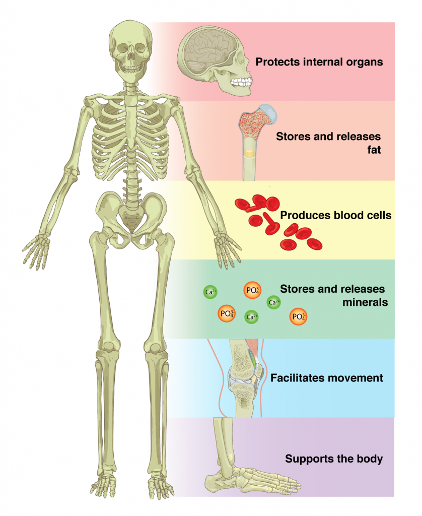
As you have already understood, your skeleton maintains a lot of different functions. Some of them, such as movement and support, were already mentioned. But let us discuss them all in detail.
- Support. Your bodies are supported by the skeleton so that you can change your position to vertical one and stand strait. Without it, you would be able only to lie because of the gravitation. This function is provided by many bones but the long ones seem to be the leaders in this competition. For instance, those that are in legs, support the trunk. Similarly, vertebras support one another so that eventually the firs one provides support to the skull. In addition to that, they support the organs and ensure that they do not change their positions.
- Protection. The skeleton also protects you. For example, the skull prevents fatal brain injuries. The rib cage protects such vital organs as the heart and lungs. It also takes care of your abdominal organs ensuring that they develop normally.
- Movement. The function of bodily motion allowed you to come here today. However, it is critical to remember that it is maintained not only due to the bones but also with the help of the muscular system.
- Mineral and energy storage. From the outer side of your bones, there is a tissue that serves as a storage. It gathers calcium and phosphorus and withdraws them to maintain appropriate blood levels. In addition to that, mature bones store yellow marrow. It consists of fat almost totally and provides you with energy for various activities.
- Blood-cell formation. The inner core of your bones takes part in the formation of blood cell and platelet. It is known as bone marrow or red marrow. Platelet is vital for you because it ensures your ability to heal wounds while blood cells spread oxygen and destroy infectious cells (CAERT 3).
Have you ever thought of the way our movement are maintained? Even a simple nod of the head requires the cooperation between the skeletal and muscular systems. Muscles ensure movement of our body through the attachment to the bones. All in all, there are about 700 of them, which is an enormous amount that comprises about 50% of your weight.
So what happens in your body when you moves? When you want to move, your brain sends a message for the body to release energy. In medical terms, it is called adenosine triphosphate. Affecting your muscles, it makes them contract or shorten. Shortened muscles pulls bones at their insertion point. Thus, the angle between the bones connected by a joint shortens. Relaxation is maintained when the opposing muscle extends and pulls a bone to its initial position.
Human skeletons seem to be similar, as they contain the same bones. However, you should remember that their characteristics differ depending on the gender. For example, women have lighter pelvis bones that form a shorter cavity with less dimensions. It has less prominent marking for muscles and more circular pelvic brim. The sacral bones of men are longer and narrower, which makes them more massive. Their femur is also longer and heavier. Its texture is rough unlike women’s smooth.
Muscle marking is more developed and shaft is less oblique. The head of men’s femur is larger and trochanters are more prominent. The femoral neck angle in males is more than 125 and in females is less than 125. Women’s sternum is less than twice the length of manubrium and larger in men. Differences in skull include greater capacity, thicker walls, more marked muscular ridges, prominent air sinuses, smoother upper margin of orbit, less vertical forehead, and heavier cheekbones in males.
Hopefully, it will never affect any of you but the skeleton may be affected by tumours that cause bone defects. People may have skeletal developmental disorders including gigantism, dwarfism, osteogenesis imperfecta, and rickets lead to abnormal body sizes, brittle bones, and growth retardation. Bacterial infections cause inflammation and lead to bone destruction.
Decalcification, including the known to you osteoporosis, reduces bone tissue and softens bones. Joint disorders often deal with inflammation. For instance, arthritis. They are often influenced by age and physical activity. In this way, degradation of joints is observed in the elderly but can be delayed due to regular exercises. The abnormal curvatures of the spine may also cause health issues. That is why you should pay attention to your back posture and avoid kyphosis (a hunchback condition), lordosis (a swayback condition), and scoliosis (an abnormal lateral curvature).
CAERT. Structures and Functions of the Skeletal System . 2014. Web.
Skeletal System: Bones and Joints. 2012. Web.
“ The Axial & Appendicular Skeleton. ” TeachPE , 2017. Web.
- Phototransduction Process and Optical Imaging
- "Cellular Metabolism and Disease" by DeBerardinis et al.
- Articular and Muscular Systems
- The Muscular System of a Human Body
- Aspects of the Skeletal System
- Neuropsychological Tests Reliability Following Concussion
- Physicians, Their Roles and Responsibilities
- Prevalence of Sleep Disorders among Medical Students
- Human Physical Performance Under Adverse Conditions
- Tongue and Why It Is Unique
- Chicago (A-D)
- Chicago (N-B)
IvyPanda. (2020, September 6). The Skeletal System. https://ivypanda.com/essays/the-skeletal-system/
"The Skeletal System." IvyPanda , 6 Sept. 2020, ivypanda.com/essays/the-skeletal-system/.
IvyPanda . (2020) 'The Skeletal System'. 6 September.
IvyPanda . 2020. "The Skeletal System." September 6, 2020. https://ivypanda.com/essays/the-skeletal-system/.
1. IvyPanda . "The Skeletal System." September 6, 2020. https://ivypanda.com/essays/the-skeletal-system/.
Bibliography
IvyPanda . "The Skeletal System." September 6, 2020. https://ivypanda.com/essays/the-skeletal-system/.
- Success stories
- Spine and back
- Pelvis and perineum
- Head and neck
- Neuroanatomy
- Cross sections
- Radiological anatomy
- Types of tissues
- Body systems

Register now and grab your free ultimate anatomy study guide!

Musculoskeletal system
Author: Gordana Sendić, MD • Reviewer: Jana Vasković, MD Last reviewed: November 03, 2023 Reading time: 28 minutes
/images/vimeo_thumbnails/258827578/ahcKVZU8g6lWyobtGVy30g_overlay.jpg)
The musculoskeletal system (locomotor system) is a human body system that provides our body with movement , stability, shape, and support. It is subdivided into two broad systems:
- Muscular system , which includes all types of muscles in the body. Skeletal muscles, in particular, are the ones that act on the body joints to produce movements. Besides muscles, the muscular system contains the tendons which attach the muscles to the bones.
- Skeletal system , whose main component is the bone . Bones articulate with each other and form the joints , providing our bodies with a hard-core, yet mobile, skeleton. The integrity and function of the bones and joints is supported by the accessory structures of the skeletal system; articular cartilage , ligaments , and bursae .
Besides its main function to provide the body with stability and mobility, the musculoskeletal system has many other functions; the skeletal part plays an important role in other homeostatic functions such as storage of minerals (e.g., calcium) and hematopoiesis, while the muscular system stores the majority of the body's carbohydrates in the form of glycogen.
This article will introduce you to the anatomy and function of the musculoskeletal system.
| Definition | A human body system that provides the body with movement, stability, shape, and support |
| Components | Muscular system: skeletal muscles and tendons Skeletal system: bones, joints; associated tissues (cartilage, ligaments, joint capsule, bursae) |
| Function | Muscles: Movement production, joint stabilization, maintaining posture, body heat production Bones: Mechanical basis for movements, providing framework for the body, vital organs protection, blood cells production, storage of minerals |
Muscular system
Muscle contraction, functions of the muscular system, functions of the skeletal system, osteoporosis, muscular dystrophy, related articles.
The muscular system is an organ system composed of specialized contractile tissue called the muscle tissue . There are three types of muscle tissue, based on which all the muscles are classified into three groups:
- Cardiac muscle , which forms the muscular layer of the heart ( myocardium )
- Smooth muscle , which comprises the walls of blood vessels and hollow organs
- Skeletal muscle , which attaches to the bones and provides voluntary movement.
Based on their histological appearance, these types are classified into striated and non-striated muscles; with the skeletal and cardiac muscles being grouped as striated , while the smooth muscle is non-striated . The skeletal muscles are the only ones that we can control by the power of our will, as they are innervated by the somatic part of the nervous system . In contrast to this, the cardiac and smooth muscles are innervated by the autonomic nervous system , thus being controlled involuntarily by the autonomic centers in our brain .
Skeletal muscles
The skeletal muscles are the main functional units of the muscular system. There are more than 600 muscles in the human body. They vary greatly in shape in size, with the smallest one being the stapedius muscle in the inner ear, and the largest one being the quadriceps femoris muscle in the thigh.
The skeletal muscles of the human body are organized into four groups for every region of the body:
- Muscles of the head and neck , which include the muscles of the facial expression , muscles of mastication , muscles of the orbit , muscles of the tongue , muscles of the pharynx , muscles of the larynx , and muscles of the neck
- Muscles of the trunk , which include the muscles of the back , anterior and lateral abdominal muscles , and muscles of the pelvic floor
- Muscles of the upper limbs , which include muscles of the shoulder , muscles of the arm , muscles of the forearm and muscles of the hand
- Muscles of the lower limbs , which include hip and thigh muscles , leg muscles and foot muscles
The fact that there are more than 600 muscles in the body can be quite intimidating. If you’re tired of all the big, comprehensive anatomy books, take a look at our condensed muscle anatomy reference charts , which contain all the muscle facts in one place organized into neat tables!
Structurally, the skeletal muscles are composed of the skeletal muscle cells which are called the myocytes (muscle fibres, or myofibrils ). Muscle fibers are specialized cells whose main feature is the ability to contract. They are elongated, cylindrical, multinucleated cells bounded by a cell membrane called sarcolemma . The cytoplasm of skeletal muscle fibers ( sarcoplasm ), contains contractile proteins called actin and myosin. These proteins are arranged into patterns, forming the units of contractile micro-apparatus called sarcomeres .
Each muscle fiber is enclosed with a loose connective tissue sheath called endomysium . Multiple muscle fibers are grouped into muscle fascicles or muscle bundles, which are encompassed by their own connective tissue sheath called the perimysium . Ultimately, a group of muscle fascicles comprises a whole muscle belly which is externally enclosed by another connective tissue layer called the epimysium . This layer is continuous with yet another layer of connective tissue called the deep fascia of skeletal muscle, that separates the muscles from other tissues and organs.
This structure gives the skeletal muscle tissue four main physiological properties:
- Excitability - the ability to detect the neural stimuli ( action potential );
- Contractibility - the ability to contract in response to a neural stimulus;
- Extensibility - the ability of a muscle to be stretched without tearing;
- Elasticity - the ability to return to its normal shape after being extended.
Learn everything about the skeletal muscle structure with our articles, video tutorials, quizzes and labelled diagrams.
:format(jpeg)/images/study_unit/skeletal-muscle-tissue/1eGzo2zCXJgncriRIxgslA_Skeletal_muscle.png "essay on skeleton system")
The most important property of skeletal muscles is its ability to contract . Muscle contraction occurs as a result of the interaction of myofibrils inside the muscle cells. This process either shortens the muscle or increases its tension, generating a force that either facilitates or slows down a movement.
There are two types of muscle contraction; isometric and isotonic. A muscle contraction is deemed as isometric if the length of the muscle does not change during the contraction, and isotonic if the tension remains unchanged while the length of the muscle changes. There are two types of isotonic contractions:
- Concentric contraction , in which the muscle shortens due to generating enough force to overcome the imposed resistance. This type of contraction serves to facilitate any noticeable movement (e.g. lifting a barbell or walking on an incline).
- Eccentric contraction , in which the muscle stretches due to the resistance being greater than the force the muscle generates. During an eccentric contraction, the muscle maintains high tension. This type of contraction usually serves to slow down a movement (e.g. lowering a barbell or walking downhill).
The sequence of events that results in the contraction of a muscle cell begins as the nervous system generates a signal called the action potential . This signal travels through motor neurons to reach the neuromuscular junction , the site of contact between the motor nerve and the muscle. A group of muscle cells innervated by the branches of a single motor nerve is called the motor unit .
The incoming action potential from the motor nerve initiates the release of acetylcholine (ACh) from the nerve into the synaptic cleft , which is the space between the nerve ending and the sarcolemma. The ACh binds to the receptors on the sarcolemma and triggers a chemical reaction in the muscle cell. This involves the release of calcium ions from the sarcoplasmic reticulum , which in turn causes a rearrangement of contractile proteins within the muscle cell. The main proteins involved are actin and myosin, which in the presence of ATP, slide over each other and pull on the ends of each muscle cell together, causing a contraction. As the nerve signal diminishes, the chemical process reverses and the muscle relaxes.
A tendon is a tough, flexible band of dense connective tissue that serves to attach skeletal muscles to bones. Tendons are found at the distal and proximal ends of muscles, binding them to the periosteum of bones at their proximal ( origin ) and distal attachment ( insertion ) on the bone. As muscles contract, the tendons transmit the mechanical force to the bones, pulling them and causing movement.
Being made of dense regular connective tissue, the tendons have an abundance of parallel collagen fibers, which provide them with high tensile strength (resistance to longitudinal force). The collagen fibers within a tendon are organized into fascicles, and individual fascicles are ensheathed by a thin layer of dense connective tissue called endotenon . In turn, groups of fascicles are ensheathed by a layer of dense irregular connective tissue called epitenon . Finally, the epitenon is encircled with a synovial sheath and attached to it by a delicate connective tissue band called mesotenon .
Learn more about the microstructure of tendon in this study unit:
:format(jpeg)/images/study_unit/dense-connective-tissue/6vZ9mKfCxlRli5PTekyGvg_Dense_connective_tissue.png "essay on skeleton system")
The main function of the muscular system is to produce movement of the body. Depending on the axis and plane, there are several different types of movements that can be performed by the musculoskeletal system. Some of the most important ones include:
- Flexion and extension : movement of decreasing or increasing the angle between the bones involved in the movement, respectively. This motion takes place in the sagittal plane around a frontal axis. An example of flexion is bending the leg at the knee joint , whereas extension would be straightening knee from a flexed position.
- Adduction and abduction : movements of bringing the parts of the body towards or away from the midline, respectively. These movements are carried out in the frontal plane around a sagittal axis. For example, abduction of the arm at the shoulder joint involves moving the arm away from the side of the body, while adduction involves bringing it back towards the body.
- Rotation is the movement in which a part of the body rotates around its vertical (longitudinal) axis in the transverse plane. This movement is defined relative to the midline, where internal rotation involves rotating the segment towards to the midline, while external rotation involves moving it away from the midline. Examples include lateral or medial rotation of the thigh .
- Supination and pronation are special types of rotatory movements usually used to describe the movements of the forearm . Supination is essentially a lateral rotation of the forearm which turns the palms anteriorly (if the arm is anatomical position) or superiorly, when the elbow is flexed. These movements are also sometimes used to describe movements in the ankle and foot , in which supination means rolling the foot outwards, while pronation means rolling the foot inwards.
:format(jpeg)/images/study_unit/body-movements/lmBBr2EccVXyhM71UrtVg_movements.png "essay on skeleton system")
Both during movement and stationary positions, muscles contribute to the overall support and stability of joints . Many muscles and their tendons pass over joints and thereby stabilize the articulating bones and hold them in position. In addition, the muscles also play an important role in maintaining posture . While the movements occur mainly due to muscles intermittently contracting and relaxing, the posture is maintained by a sustained tonic contraction of postural muscles. These muscles act against gravity and stabilize the body during standing or walking. The postural muscles include the muscles of the back and abdominal muscles.
Another important function of muscles is heat production . Muscle tissue is one of the most metabolically active tissues in the body, in which approximately 85 percent of the heat produced in the body is the result of muscle contraction. This makes the muscles essential for maintaining normal body temperature.
Wondering what’s the best way to learn and understand the functional anatomy of the muscles? Check out our 3D muscle anatomy videos !
To improve your understanding of muscular system terminology, take a closer look at some commonly used roots, prefixes and suffixes related to the muscular system in the video below.
How well do you know the main muscles of the body? Test your knowledge with our quiz in different difficulty levels!
Skeletal system
The adult human skeleton is composed of 206 bones and their associated cartilages. The bones are supported by ligaments, tendons, bursae, and muscles. The bones of the body are grouped within the two distinct divisions:
- Axial skeleton , that includes the bones along the long axis of the body. The axial skeleton consists of the vertebral column , bones of the head and bones of the thoracic cage .
- Appendicular skeleton , that involves the bones of the shoulder and pelvic girdle , as well as the bones of the upper and lower extremities .
:format(jpeg)/images/study_unit/bones-skeletal-system/bX3KgmwVAC3JsO181kB6w_Skeletal_system.png "essay on skeleton system")
Bones are rigid structures made of calcified dense connective tissue. Bone tissue is composed of a mineralized bone matrix that consists of type 1 collagen fibers dispersed throughout the ground substance . The cellular component of the bones is represented by three types of specialized bone cells called osteocytes, osteoblasts and osteoclasts .
The bones consist of two distinct layers that differ in histological appearance and characteristics;
- Compact (cortical) bone is the outer much denser layer of the bone which gives it its smooth, white, and solid appearance. The outer surface of the compact bone is covered with a layer of dense connective tissue called the periosteum. On its inner surface, the compact bone is covered with endosteum , which is the boundary between the compact and spongy bones .
- Spongy (cancellous) bone is the deep airy layer of the bone. Unlike the compact bone, spongy bone is highly vascularized and more metabolically active. It is typically found within the ends of long bones and in the vertebrae. In certain bones, like the hip bone , sternum or femur, the central part of spongy bone houses the bone marrow, which is the site of hematopoiesis in the adult.
:format(jpeg)/images/study_unit/histology-bone-tissue/vmC3Uigzl0S5xRQahVyXNA_bone.png "essay on skeleton system")
Types of bones
Bones can be classified according to their shapes as follows:
- Long bones have a tubular shape, with a longer longitudinal and a shorter transverse diameter. They are composed mostly of compact bone, while the spongy bone and bony marrow fill the ends of the bones. Examples of long bones include the humerus , ulna , tibia and clavicle .
- Short bones have a roughly cuboid or round shape, and only contain a thin layer of compact bone surrounding the spongy bone. Examples include the tarsal and carpal bones .
- Flat bones are mostly thin, flattened and usually curved. They contain two parallel layers of compact bones surrounding a layer of spongy bone. Examples include most of the skull bones , scapula , sternum and sacrum .
- Sesamoid bones are small, rounded unique types of bones that are embedded in muscle tendons where the tendon passes over a joint. The largest sesamoid bone in the body is the patella , but several other smaller sesamoid bones can be found in the hand and foot, usually in close proximity to the joints.
- Irregular bones do not fit into any of the other categories. Generally, irregular bones contain foramina through which soft tissue and neurovascular structures pass. Examples include the vertebrae , hip bone and some bones of the skull.
Wondering how to cut time in learning the bones of the body? Try our skeletal system quizzes !
A typical long bone consists of a long shaft ( diaphysis ) that extends into a neck ( metaphysis ) and head ( epiphysis ) on its proximal and distal ends. It also features various markings and formations that give passage to neurovascular structures, as well as the attachment sites to the ligaments and tendons. Some of those features include:
- Sulcus – a shallow groove on the bone surface (e.g. radial sulcus of humerus)
- Condyle – rounded articular area (e.g. lateral condyle of tibia)
- Epicondyle – eminence superior to a condyle (medial epicondyle of femur)
- Crest – ridge of bone (e.g. iliac crest)
- Facet – smooth, flat area, usually covered with cartilage (e.g. articular facet on vertebrae)
- Foramen – passage through a bone (e.g. foramen magnum on the occipital bone)
Cartilage is a flexible connective tissue found in multiple organ systems of the body. Cartilage is composed of specialized cells called chondrocytes , collagen fibers and abundant ground substance rich in proteoglycan and elastin fibers.
Cartilage is classified into the following types based on its composition:
- Hyaline cartilage is composed of type II collagen and an abundance of ground substance, which gives it a glossy appearance. It is the most abundant type of cartilage found in joints (articular cartilage), as well as the nose, larynx , trachea and ribs .
- Elastic cartilage is similar to hyaline cartilage but contains more elastic fibers. It is found in structures such as the pinna of the ear , auditory tube and epiglottis .
- Fibrocartilage is composed of plenty of collagen fibers type I and a smaller amount of ground substance. Examples of fibrocartilage include intervertebral discs , pubic and other symphyses.
The musculoskeletal system specifically contains articular cartilage, a type of cartilage that lines the articulating surfaces of bones. The articular cartilage provides congruence to the articulating bones and allows them to bear weight and glide over each other with very little friction.
:format(jpeg)/images/study_unit/histology-hyaline-cartilage/4Sc33EqhMjeN76FFmhpDQ_Hyaline_cartilage.png "essay on skeleton system")
Each bone of the musculoskeletal system is connected to one or more bones via a joint . Joints provide a fulcrum to the bones, on which they pivot and thereby allow movements of body parts. However, movement is not a necessary attribute of a joint as some joints do not move, such as joints between the bones of the skull. The integrity or stability of a joint is provided by several factors including the bony congruence and structures that cross the joint, such as tendons and ligaments.
Based on the type of tissue that holds the neighboring bones together and the range of motion they exhibit, joints can be classified into the following:
- Synovial joints are freely mobile joints in which the bones are not in direct contact, but are separated by a potential space called the synovial cavity . The synovial cavity is lined by a synovial membrane that secretes the synovial fluid which nourishes and lubricates the articulating surfaces in order to reduce friction. The articulating bones in most synovial joints are lined with hyaline cartilage. These joints usually have a wide range of motion, which is defined by the joint capsule, the supporting ligaments and muscles that cross the joint. Examples of synovial joints include the knee, shoulder, sternoclavicular and elbow joints .
- Fibrous joints are the articulations in which the bones are connected by dense fibrous connective tissue. The bones in fibrous joints are firmly held together so that the joint allows negligible movement. Fibrous joints are found between the cranial sutures , the distal tibiofibular and cuboideonavicular joints.
- Cartilaginous joints are articulations in which the bones are connected by cartilage. The bones have a range of motion between synovial and fibrous joints. Cartilaginous joints are subdivided into synchondrosis (e.g. costochondral joints ) and symphysis joints (e.g. pubic symphysis).
According to the movements they allow and/or the shape of their articulating surface, the synovial joints can be further subdivided into 6 major types:
- Ball and socket joints (e.g. hip joint )
- Condyloid joints (e.g. metacarpophalangeal joint)
- Hinge joints (e.g. elbow joint)
- Pivot joints (e.g. atlanto-axial joint)
- Saddle joints (e.g. carpometacarpal joint)
- Plane joints (e.g. acromioclavicular joint )
Ligaments are fibrous bands made of dense regular connective tissue which are similar in structure to tendons. Unlike the tendons that connect muscles to bone, the ligaments connect bone to bone . Besides the musculoskeletal system, the ligaments are also found in many other parts of the body, where they usually stabilize and hold internal organs in place and transmit neurovascular structures.
In the musculoskeletal system, ligaments stabilize the articulating bones and reinforce the joints. Depending on their anatomic position relative to the joint capsule, ligaments are classified into:
- Capsular ligaments are essentially thickenings of the joint capsule that form either elongated bands or triangular structures. These ligaments serve to reinforce the integrity of the joint capsule. An example of the capsular ligament is the iliofemoral ligament of the hip joint.
- Intracapsular ligaments are the ligaments that lie internal to the joint capsule. These ligaments reinforce the connection of the articulating surfaces of the joint, but allow a far wider range of motion than other ligaments. Examples include anterior and posterior cruciate ligament of the knee joint.
- Extracapsular ligaments are ligaments that lie outside the joint capsule. These ligaments provide the most stability to the articulating bones, and are important for preventing dislocations. Extracapsular ligaments can lie in close proximity (e.g. medial collateral ligament of the ankle joint ) or a bit further from the joint capsule ( vertebral ligaments ).
Bursae are small sac-like outpouchings of the joint cavity lined by synovial membrane. They are found around the joints, providing cushioning of the associated bones, tendons and muscles and reducing friction between adjacent structures.
The majority of synovial bursae are located near the large joints of the arms and legs. For example, one of the bursae of the knee joint is the suprapatellar bursa , found superior to the patella, between the femur and the tendon of the quadriceps femoris muscle . The suprapatellar bursa allows for these structures to slide over each other without friction during flexion and extension of the knee joint.
To improve your understanding of skeletal system terminology, take a closer look at some commonly used roots, prefixes and suffixes related to the skeletal system in the video below.
Time for a skeletal system workout with our integrated quiz!
The skeletal system serves a variety of functions. The bones give the shape to the body and provide the site of attachment to muscles, tendons, ligaments and cartilage. These tissues function together as a whole to generate a force that provides the biomechanical basis of movement .
Due to its structural integrity, the skeletal system protects the internal organs, most importantly the brain, which is surrounded by the skull, as well as the heart and lungs , which are protected by the rib cage.
Moreover, the skeletal system serves several metabolic functions . The bones are the storage site of important minerals, most notably calcium and phosphorus. This makes the bones essential for balancing calcium levels in the blood, which is regulated by adjusting the rate of bone resorption.
Lastly, the bone marrow found in spongy bone is the site of hematopoiesis , which is a process of production of new blood cells. Cells that are produced in the bone marrow are red blood cells, platelets and white blood cells, such as monocytes, granulocytes and lymphocytes .
We created a custom summary quiz about the anatomy and histology of the main features of the musculoskeletal system. You can adjust and filter individual structures to make this quiz your own!

Clinical correlation
There is a variety of conditions that affect the muscles, bones, and joints. Disorders of the musculoskeletal system may range from diseases to minor physical disabilities. The following are some clinical conditions of the musculoskeletal system:
Osteoporosis is a condition that affects bone strength (the word osteoporosis literally means "porous bones"). It is a condition in which the bones become fragile and brittle, leading to a higher risk of fractures than in normal bone. As a result, even a minor bump or accident can cause serious fractures.
Osteoporosis is the “bone of the old”, especially, in women. The hard, rock-like quality of bone is dependent upon calcium. When too much calcium is dissolved from bones or not enough is replaced, bones lose density and are easily fractured. Estrogen, the female sex hormone, helps maintain proper calcium levels in bones. Once the ovaries stop producing the hormone, women are at higher risk of developing osteoporosis. A collapse of bony vertebrae of the spinal column results in loss of height and stooped posture. Hip fractures are a common occurrence.
Sarcopenia is a syndrome characterized by progressive and generalized loss of skeletal muscle mass and strength with a risk of adverse outcomes such as physical disability, poor quality of life and death.
Arthritis is a group of conditions affecting the joints . These conditions cause damage to the joints, usually resulting in pain and stiffness due to aging. Arthritis can affect many different parts of the joint and nearly every joint in the body.
As an individual ages, the joint tissues become less resilient to wear and tear and start to degenerate. This degeneration manifest as swelling, pain, and often-times, loss of mobility of joints. Changes occur in both joint soft tissues and the articulating bones, a condition called osteoarthritis . A more serious form of disease is called rheumatoid arthritis . The latter is an autoimmune disease wherein the body produces antibodies against joint tissues causing chronic inflammation resulting in severe joint damage, pain and immobility.
Muscular dystrophy is a group of muscle diseases that weaken the musculoskeletal system and hamper locomotion. Muscular dystrophies are characterized by progressive skeletal muscle weakness , defects in muscle proteins, and the death of muscle fibres (muscle cells) and tissue.
It is a group of inherited diseases in which the muscles that control movement progressively weaken. The prefix, dys-, means abnormal, while the root, -trophy, refers to maintaining normal nourishment, structure and function. The most common form in children is called Duchenne muscular dystrophy and affects only males. It usually appears between the ages of 2 to 6 and the afflicted live typically into late teens to early 20s.
Other conditions involving the musculoskeletal system include:
- Lupus erythematosus
- Myasthenia gravis
- Rotator cuff tear
- Carpal tunnel syndrome
- Osteomalacia
References:
- Moore, K. L., Dalley, A. F., & Agur, A. M. R. (2014). Clinically Oriented Anatomy (7th ed.). Philadelphia, PA: Lippincott Williams & Wilkins.
- Netter, F. (2019). Atlas of Human Anatomy (7th ed.). Philadelphia, PA: Saunders.
- Standring, S. (2016). Gray's Anatomy (41st ed.). Edinburgh: Elsevier Churchill Livingstone.
- Ross, Lawrence M; Lamperti, Edward D, eds. (2006). Thieme Atlas of Anatomy: General Anatomy and Musculoskeletal System.
- R.M.H McMinn: Last's anatomy (Regional and Applied), 9th edition, Ana-Maria Dulea (2014
Illustrations:
- Musculoskeletal system - Irina Münstermann
- Eccentric and concentric muscle contractions (diagram) - Yousun Koh
Articles within this topic:
- Ball and socket joint
- Complete list of bone markings
- Ellipsoid joint
- Head and neck anatomy
- Hinge-joint
- How to learn all muscles with quizzes and labeled diagrams
- Intercostal muscles
- Lateral abdominal muscles
- Learn skull anatomy with skull bones quizzes and diagrams
- Lower limb anatomy
- Muscle anatomy reference charts
- Muscles of the neck: An overview
- Muscles of the trunk
- Musculoskeletal system development
- Parietal bone
- Pectoralis minor muscle
- Pivot joint
- Rectus abdominis muscle
- Synovial membrane
- Upper limb muscles and movements
Musculoskeletal system: want to learn more about it?
Our engaging videos, interactive quizzes, in-depth articles and HD atlas are here to get you top results faster.
What do you prefer to learn with?
“I would honestly say that Kenhub cut my study time in half.” – Read more.

Learning anatomy isn't impossible. We're here to help.

Learning anatomy is a massive undertaking, and we're here to help you pass with flying colours.
Want access to this video?
- Curated learning paths created by our anatomy experts
- 1000s of high quality anatomy illustrations and articles
- Free 60 minute trial of Kenhub Premium!
...it takes less than 60 seconds!
Want access to this quiz?
Want access to this gallery.
- Type 2 Diabetes
- Heart Disease
- Digestive Health
- Multiple Sclerosis
- Diet & Nutrition
- Supplements
- Health Insurance
- Public Health
- Patient Rights
- Caregivers & Loved Ones
- End of Life Concerns
- Health News
- Thyroid Test Analyzer
- Doctor Discussion Guides
- Hemoglobin A1c Test Analyzer
- Lipid Test Analyzer
- Complete Blood Count (CBC) Analyzer
- What to Buy
- Editorial Process
- Meet Our Medical Expert Board
The Anatomy of the Skeletal System
The skeletal system comprises 206 bones and has two main parts—the axial skeleton and the appendicular skeleton. The skeletal system includes your bones, ligaments that attach bone to bone, and cartilage that provides padding between your bones.
This article discusses the anatomy of the skeletal system—what it's made of, how it's organized, conditions that affect it, and tests that assess it.
SDI Productions / Getty Imgaes
Skeletal System: Labeled Diagram of Major Organs
In addition to the bones, organs of the skeletal system include ligaments that attach bones to other bones and cartilage that provides padding between bones that form joints throughout your body.
The bones are divided into two main categories—the axial skeleton , which contains the bones that support the middle of your body, and the a ppendicular skeleton, which includes bones that make up your appendages—arms and legs—and bones that attach your limbs to your axial skeleton.
Axial Skeleton
The axial skeleton forms the "axis" that runs down the center of the body. There are 80 bones that make up the axial skeleton.
Skull (Cranium)
Your skull ( cranium ) is made up of cranial and facial bones. Cranial bones protect your brain, while facial bones make up your facial structure. Skull bones include the following:
Cranial bones include:
Facial bones include:
- Maxillae (upper jaw)
- Mandible (lower jaw)
- Zygomatic (cheekbones)
- Inferior nasal conchae
Auditory Ossicles
The auditory ossicles consist of a total of six tiny bones, with three in each ear. They are located in the inner ear and are structures that help create sound in your body. The auditory ossicles include the following:
The hyoid bone is a horseshoe-shaped bone located in the throat. It is part of bodily functions like speaking, swallowing, and airway maintenance.
Vertebral column
The vertebral column (spine) protects your spinal cord, supports your head, and allows bodily movement. It contains the sacrum (made up of four bones) and coccyx (the tailbone, which is made up of five bones), and 24 vertebrae, including:
- Cervical vertebrae : Seven bones in the neck region
- Thoracic vertebrae : Twelve bones attached to the ribs
- Lumbar vertebrae : Five bones in the low back region
The thorax contains the sternum (breastbone) and the thoracic (rib) cage . The thoracic cage comprises 12 pairs of ribs connecting to the thoracic vertebrae and the sternum. Your rib cage protects your heart.
Appendicular Skeleton
The appendicular skeleton includes 126 bones that comprise your appendages—your arms and legs—and the bones that attach your limbs to your axial skeleton.
Upper Extremities
Your upper extremities refer to your shoulders and arms. Bones in the upper extremities include:
- Scapula (shoulder blade)
- Clavicle (collarbone)
- Humerus (upper arm)
- Radius and ulna (forearm bones)
- Carpals (eight tiny bones in the wrist)
- Metacarpals (in the palm)
- Phalanges (bones of the fingers)
Lower Extremities
Bones in the lower extremities make up your hips and legs and include:
- Femur (thigh bone)
- Patella (kneecap)
- Tibia and fibula (lower leg bones)
- Tarsals (eight tiny bones in the ankle)
- Metatarsals (in the middle of the foot)
- Phalanges (bones of the toes)
Which Bones Are Most Commonly Broken?
The most commonly fractured bones include the distal radius (on the thumb side of your wrist), the ankle, the femur (thigh bone), the humerus (upper arm bone), and the metacarpals (bones of the palms).
With osteoporosis, the most commonly fractured bones are the vertebrae (in the spine).
What Is the Purpose of the Skeletal System?
The primary purpose of the skeletal system is to give the body its shape and to provide attachment points for the muscles that move the body.
Other purposes of the skeletal system include:
- Storing minerals (such as calcium) and fats
- Producing red blood cells
- Protecting internal organs
Calcium in Your Bones
Most of the body's calcium is stored in your bones.
Skeletal System Associated Conditions
Various conditions and injuries can affect the skeletal system. Examples include:
- Fractures (broken bones)
- Ligament sprains
- Osteoarthritis
- Osteoporosis
- Osteogenesis imperfecta
- Osteomalacia
- Paget's disease of bone
- Osteomyelitis
- Avascular necrosis (osteonecrosis)
- Marfan syndrome
- Ehlers-Danlos syndrome
- Osteopetrosis
- Ankylosing spondylitis
- Bone marrow diseases
Skeletal System Tests
Many different tests can help diagnose conditions that affect the skeletal system.
Imaging Tests
Healthcare providers use various imaging tools to get detailed pictures of your bones. Depending on the reason for imaging, a healthcare provider will conduct one or more of the following tests:
- X-rays : This common test can help diagnose conditions that affect the bones and joints, such as fractures or arthritis.
- Computed tomography (CT scan), computerized axial tomography (CAT scan) : This test provides three-dimensional pictures that help diagnose fractures that aren't clear on X-rays, or other bone conditions, such as cancer.
- Magnetic resonance imaging (MRI) : This type of imaging often helps diagnose conditions that affect soft tissues of the skeletal system (ligaments, cartilage).
- Bone scintigraphy (bone scan) : These scans can provide detailed information about a bone injury or condition, such as the staging of bone cancer.
- Positron emission tomography (PET scan) : This test, which uses an injected radioactive tracer, can help stage bone (and other types of) cancer.
- Bone density test : These tests are primarily for determining how dense bones are—the key factor in diagnosing osteoporosis.
Other Skeletal System Tests
Healthcare providers may perform additional tests if they need further information about your bones or skeletal system. These tests may include:
- Joint aspiration : This test involves removing a sample of fluid from a joint to help diagnose infection.
- Biopsy : For the skeletal system, this procedure can involve removing a small sample of bone or bone marrow so that it can be tested for conditions such as cancer.
- Blood tests : These tests help diagnose infections that can affect the skeletal system.
The skeletal system is made up of your bones, ligaments, and cartilage. Though its main function is to provide structural support for the body, it also stores important minerals—such as calcium—forms red blood cells, and protects your internal organs. The skeletal system can break down into two main categories—the axial skeleton, which forms the "long axis" of the body, and the appendicular skeleton, which forms your arms and legs.
Many different injuries and diseases can affect the skeletal system. Imaging procedures, such as X-rays, MRI, and bone density tests, and other tests, such as blood work or tissue biopsy, can help to diagnose these conditions.
National Cancer Institute. Cranium .
National Cancer Institute SEER Training Modules. Axial skeleton (80 bones) .
Fisher E, Austin D, Werner HM, et al. Hyoid bone fusion and bone density across the lifespan: prediction of age and sex . Forensic Sci Med Pathol . 2016;12(2):146-157. doi:10.1007/s12024-016-9769-x
Osmosis from Elsevier. Bones of the vertebral column .
National Cancer Institute SEER Training Modules. Appendicular skeleton (126 bones) .
American Academy of Orthopaedic Surgeons. Osteoporosis and spinal fractures .
National Cancer Institute SEER Training Modules. Introduction to the skeletal system .
MedlinePlus. Calcium and bones .
National Institute of Arthritis and Musculoskeletal and Skin Diseases. Muscle and bone diseases .
MedlinePlus. Diagnostic imaging .
InformedHealth.org. Understanding tests used to detect bone problems .
Johns Hopkins Medicine. Joint aspiration .
By Aubrey Bailey, PT, DPT, CHT Dr, Bailey is a Virginia-based physical therapist and professor of anatomy and physiology with over a decade of experience.
- COVID-19 Tracker
- Biochemistry
- Anatomy & Physiology
- Microbiology
- Neuroscience
- Animal Kingdom
- NGSS High School
- Latest News
- Editors’ Picks
- Weekly Digest
- Quotes about Biology
Skeletal System
Reviewed by: BD Editors
The skeletal system provides support and protection for the body’s internal organs and gives the muscles a point of attachment. Humans have an endoskeleton , where our bones lie underneath our skin and muscles. In other animals, such as insects, there is an exoskeleton on the outside of the body.
In humans, the skeletal system consists of bones, joints and associated cartilages. An adult human has 206 bones in their body and variety of different joints.

Image shows a human skeleton with the major bones labeled.
The human skeleton can be divided into two components: the axial skeleton and the appendicular skeleton. The axial skeleton is formed around the central axis of the body and thus includes the skull, spine , and ribcage. It protects the brain, spinal cord, heart, lungs, esophagus and major sense organs like the eyes, ears, nose, and tongue. The appendicular skeleton is related to the limbs and consists of the bones of the arms and legs, as well as the shoulder and hip girdles.
Skeletal System Function
The first and most apparent function of the skeletal system is to provide a framework for the body. The presence of a firm bony skeleton allows the organism to have a distinctive shape adapted towards a particular lifestyle. For instance, in a fast-moving animal like the cheetah, the skeleton contains long, thin limb bones and an extremely flexible spine. The structure of the skeleton also allows it to absorb the impact of running at high speeds.
The bones of birds are hollow, light and create a streamlined body adapted for flight. Many animals even have sexual dimorphism in their skeletons. In humans, while this dimorphism is fairly limited, there are differences in the angle of the pelvic bones, to accommodate pregnancy.
Integration with the Muscular System
The skeletal system also provides an important form of attachments to the muscular system. Bones and exoskeletons are hard and do not bend or move when muscles are flexed. This means that the contraction of muscle cells will lead to the shortening of muscles, while the bone retains its shape. This basic structure allows muscles to move different parts of the body, using forces generated while pulling on the skeletal system.
The next obvious function of the skeletal system is the role it plays protecting the fragile internal organs. In humans, this is seen in the skull, which surrounds the brain completely. It is also exhibited by the ribcage, which surrounds the lungs and heart but still allows for expansion. Even invertebrates like snails and prawns often have hard exoskeletons to protect themselves from predators.
The rigid endoskeleton allows the body to rise up above the ground or stand upright, and bears the weight of the organism, and provides the scaffolding for movement. Muscles generate the force required to move bones at joints. Muscle fibers contain actin and myosin, two protein filaments that can slide past each other to change the length of the muscle. When a nerve impulse arrives at the neuromuscular junction, it signals the muscle to contract. The force generated by the contracting muscle either pulls two bones together or apart, based on the nature of the interaction between the muscle and joint.
Blood Cell Production
The central part of a bone contains the bone marrow , the primary site for blood cell production in adult humans. There are two types of bone marrow in adults. Around 50% is red bone marrow containing hematopoietic stem cells and supportive tissue. The rest is yellow bone marrow made of fat and its proportion increases with age.
Bone marrow will revert to a higher proportion of red marrow if the body suffers an injury and needs to create more red blood cells. The bone marrow composition also changes during pregnancy and lactation in mammals. Over the course of gestation, blood volume increases by about 1.5 liters, and even the concentration of red blood cells and white blood cells increase.
Production of other Cell Types
In addition to producing red blood cells, bone marrow within the skeletal system is the production site of a number of other cells. These include lymphocytes , which are immune cells that travel the lymphatic system. In addition to providing immune functions, the skeletal system is also responsible for hosting stem cells which can differentiate into muscle cells, cartilage-producing cells, and cells that create bone (osteoblasts).
Osteoblasts in bone also have an endocrine function, secreting a hormone called osteocalcin. It requires vitamin K to be synthesized and is an anabolic hormone. It mediates an increase in insulin levels and increases the sensitivity of the body to insulin. Osteocalcin contributes to an increase in bone mass and bone mineralization.
Storing Minerals
The bones of the skeletal system act as a storehouse for calcium ions , changing the quantum of mineralized deposits within bones to maintain plasma calcium ion concentration within a narrow range. Calcium ions can affect crucial sodium ion channels in the plasma membrane of every cell, thereby affecting overall homeostasis.
For this reason, changes to the concentration of calcium ions have particularly adverse effects on excitable cells in the nervous system, and in cardiac, skeletal and smooth muscle. Different interacting hormones maintain the balance of calcium ions in the plasma and bones, especially the parathyroid hormone secreted from the parathyroid glands in the neck.
Skeletal System Parts
The anatomy of the skeletal system is complex, and it includes hundreds of bones in the human body . The anatomy of the system varies widely between organisms, as evolution has selected for various adaptations in certain species which change the structure and function of their bones.
Bones serve a variety of functions, but the most important is supporting movement of the limbs and body. Two bones or cartilages are held together at a joint through tough connective tissues called ligaments. Muscles are securely attached to bones through flexible but inelastic connective tissue called tendons. Muscles, joints, tendons, and ligaments are part of the intricate machinery that allows the movement of different bones.
Functionally, joints can be divided into three classes based on the range of movement they allow in the associated bones. Immovable joints are formed when two bones are held together by fibrous connective tissue with no synovial fluid. These kinds of joints hold the bones of the cranium together.
Partially movable joints are also called cartilaginous joints and are present in the spine and ribs. The third type of joints are called synovial joints and have a fluid-filled synovial cavity that allows the interfacing bones the largest range of movement. Based on the structure of the synovial joints, they can be classified into 6 types, including the hinge joints of the fingers and the ball and socket joints of the hips and shoulders.
Cellular Composition
Each bone is made of complex sets of cells, tissues and a specialized extracellular matrix . The two main cell types are called osteoblasts and osteoclasts with mostly opposing functions. While osteoblasts are involved in the formation of bone, osteoclasts are associated with a reduction in bone mass. The extracellular matrix of the bone consists of collagen and other organic fibers as well as the inorganic component containing calcium salts such as hydroxyapatite. In the interior of bones, a soft tissue called the bone marrow plays an important role in immunity and hematopoiesis. The bone is also richly supplied with nerves and blood vessels.
Skeletal System Structure
In general, the skeletal system is structured to provide support against gravity and protect an animal’s internal organs. While this article mainly discusses the human skeletal system, most animals have some sort of skeleton. Some animals, like sponges, can have an extremely simplified skeleton made of calcium deposits within the animal. Others, like the turtle, have drastically modified their skeletal system to provide extra protection.
While this article mostly discusses an endoskeleton, many animals use an exoskeleton for the same purposes. Instead of bones being on the inside, the bones, protective plates, or chitinous skeleton actually surrounds the muscles. While this may seem completely different, the structure of the system is still very similar. The only difference is that muscles and tendons connect to the inside of the system, rather than to the surface of bones.
The structure of the skeletal system reflects an animal’s evolution, as well as the needs it has to survive. For example, humans have a tailbone. This is an evolutionary relic, from the time when our ancestors had tails and were swinging from the trees. As we became bipedal, we lost the need for a tail, and it was reduced to a single, nonfunctional bone. Likewise, all animals are constantly adapting and changing their skeletal system through evolutionary time.

Skeletal System Diseases
Diseases of the skeletal system could be confined to one section of the skeleton such as changes to the curvature of the spine, or they could be a genetic disorder affecting all bones and joints such as arthritis or osteoporosis.
The spine in healthy individuals is S-shaped, with a convex curvature for the thoracic region and the concave tilt in the cervical and lumbar regions. This shape for the spine is ideally suited for an upright walking posture. If either the thoracic or lumbar regions have a change in curvature or there is sideways bend to the spine, it can lead to back pain, difficulty with breathing, digestion, mobility, and reproduction.
Curvature of the Spine
The bulk of the weight of the upper body is transmitted along the central axis towards the legs. When the bones or muscles of the back or not functioning optimally, it can lead initially to accommodative changes in posture and thereafter to pain, injury or permanent deformity. Since the spine surrounds the spinal cord, abnormalities in the skeletal structure of the spine can affect the nervous system, either manifesting as pain, tingling or numbing in the extremities. Additionally, the spine supports the ribcage, enclosing the heart, lungs, and diaphragm. Thus, spinal deformities can also lead to shortness of breath, palpitations or even cardiac arrhythmias.
Kyphosis is the term for the convex curve of the thoracic region and excessive curvature in this region is called hyperkyphosis. Extreme hyperkyphosis presents as a hunchback. This could arise from genetic factors or poor posture due to obesity or osteoporosis or arthritis.
The normal concave structure of the lumbar region is called lordosis , and when the region is overly arched, it is called lumbar hyperlordosis. In hyperlordosis, shoulders appear to be pushed back, while the abdominal region seems to be jutting forward.

Image shows a person with hyperlordosis. In a healthy spine, the midpoint of the spine (A) would be directly above the knee (B).
Hyperlordosis can arise from genetic factors, poor posture, or even deficient muscle strength. When the spine has a sideways tilt, or a lateral bend, it is called scoliosis and could be associated with both hyperkyphosis and hyperlordosis.
Osteoporosis
Osteoporosis is a condition marked by bone resorption. This reduces bone mass and density, thereby enhancing the probability of fractures from even minor stressors such as sneezing. Although osteoporosis is commonly associated with aging, smoking, obesity, diet, some medications and alcohol consumption can contribute to the progression of the ailment.
Weight training, exercise, and a diet containing adequate calcium, iron, phosphorous as well as Vitamin D, help in enhancing bone density and bone mass. There is some evidence that the pH of blood plays a role in the release of calcium stores from bones and the extent of bone mineralization since calcium salts are often used as buffers in acidic environments in the body. A whole-foods, plant-based diet has been shown greatly reduce blood acidification. As a result, it also lowers cases of osteoporosis.
Arthritis includes a number of joint disorders that are characterized by stiffness, inflammation, and pain. While there is a range of potential causes, arthritis usually worsens with age, affecting the joints that are used most frequently – especially the joints in fingers, hips, and knees. Arthritis, therefore, causes disability, restricts movement and impairs fine motor skills.
Interesting Facts
- Three bones in the inner ear, called malleus, incus and stapes, are the smallest bones in the human body. The thigh bone, or femur, is the largest bone.
- The hyoid bone situated behind the lower jaw is also called a ‘floating’ bone because it is not part of any joint, and is not directly attached to any other bone.
- The position of the hyoid bone makes it extremely resistant to fractures. However, autopsies that reveal a broken hyoid bone indicate death from strangulation.
- Newborn babies have about 300 bones. Many of these bones fuse together to form the 206 bones of the adult.
- Teeth are part of the skeletal system. However, they are not bones.
1. Which of these is an inflammatory condition?
2. Meat, carbonated beverages, cheese, eggs and even milk have been linked to a decrease in blood pH. How might these things affect bone health?
3. Why is the hyoid bone called a floating bone?
4. Why do babies have more bones than adults?
5. Which of the following are functions of the skeletal system?
Enter your email to receive results:
Cite This Article
Subscribe to our newsletter, privacy policy, terms of service, scholarship, latest posts, white blood cell, t cell immunity, satellite cells, embryonic stem cells, popular topics, mitochondria, natural selection, translation.
6.1 The Functions of the Skeletal System
Learning objectives.
By the end of this section, you will be able to:
List and describe the functions of the skeletal system
- Attribute specific functions of the skeletal system to specific components or structures
The skeletal system is the body system composed of bones, cartilages, ligaments and other tissues that perform essential functions for the human body. Bone tissue, or osseous tissue , is a hard, dense connective tissue that forms most of the adult skeleton, the internal support structure of the body. In the areas of the skeleton where whole bones move against each other (for example, joints like the shoulder or between the bones of the spine), cartilages, a semi-rigid form of connective tissue, provide flexibility and smooth surfaces for movement. Additionally, ligaments composed of dense connective tissue surround these joints, tying skeletal elements together (a ligament is the dense connective tissue that connect bones to other bones). Together, they perform the following functions:
Support, Movement, and Protection
Some functions of the skeletal system are more readily observable than others. When you move you can feel how your bones support you, facilitate your movement, and protect the soft organs of your body. Just as the steel beams of a building provide a scaffold to support its weight, the bones and cartilages of your skeletal system compose the scaffold that supports the rest of your body. Without the skeletal system, you would be a limp mass of organs, muscle, and skin. Bones facilitate movement by serving as points of attachment for your muscles. Bones also protect internal organs from injury by covering or surrounding them. For example, your ribs protect your lungs and heart, the bones of your vertebral column (spine) protect your spinal cord, and the bones of your cranium (skull) protect your brain (see Figure 6.1.1 ).
Mineral and Fat Storage, Blood Cell Formation
On a metabolic level, bone tissue performs several critical functions. For one, the bone tissue acts as a reservoir for a number of minerals important to the functioning of the body, especially calcium, and phosphorus. These minerals, incorporated into bone tissue, can be released back into the bloodstream to maintain levels needed to support physiological processes. Calcium ions, for example, are essential for muscle contractions and are involved in the transmission of nerve impulses.
Bones also serve as a site for fat storage and blood cell production. The unique connective tissue that fills the interior of most bones is referred to as bone marrow . There are two types of bone marrow: yellow bone marrow and red bone marrow. Yellow bone marrow contains adipose tissue, and the triglycerides stored in the adipocytes of this tissue can be released to serve as a source of energy for other tissues of the body. Red bone marrow is where the production of blood cells (named hematopoiesis, hemato- = “blood”, -poiesis = “to make”) takes place. Red blood cells, white blood cells, and platelets are all produced in the red bone marrow. As we age, the distribution of red and yellow bone marrow changes as seen in the figure ( Figure 6.1.2 ).
Career Connection – Orthopedist
An orthopedist is a doctor who specializes in diagnosing and treating disorders and injuries related to the musculoskeletal system. Some orthopedic problems can be treated with medications, exercises, braces, and other devices, but others may be best treated with surgery ( Figure 6.1.3 ).

While the origin of the word “orthopedics” (ortho- = “straight”; paed- = “child”), literally means “straightening of the child,” orthopedists can have patients who range from pediatric to geriatric. In recent years, orthopedists have even performed prenatal surgery to correct spina bifida, a congenital defect in which the neural canal in the spine of the fetus fails to close completely during embryologic development.
Orthopedists commonly treat bone and joint injuries but they also treat other bone conditions including curvature of the spine. Lateral curvatures (scoliosis) can be severe enough to slip under the shoulder blade (scapula) forcing it up as a hump. Spinal curvatures can also be excessive dorsoventrally (kyphosis) causing a hunch back and thoracic compression. These curvatures often appear in preteens as the result of poor posture, abnormal growth, or indeterminate causes. Mostly, they are readily treated by orthopedists. As people age, accumulated spinal column injuries and diseases like osteoporosis can also lead to curvatures of the spine, hence the stooping you sometimes see in the elderly.
Some orthopedists sub-specialize in sports medicine, which addresses both simple injuries, such as a sprained ankle, and complex injuries, such as a torn rotator cuff in the shoulder. Treatment can range from exercise to surgery.
Section Review
The major functions of the skeletal system are body support, facilitation of movement, protection of internal organs, storage of minerals and fat, and blood cell formation.
Review Questions
Critical thinking questions.
- Suppose your red bone marrow could not be formed. What functions would your body not be able to perform?
- Suppose your osseous tissue could not store calcium. What functions would your body not be able to perform?
Answers for Critical Thinking Questions
- Without red bone marrow, you would not be able to produce blood cells. The red bone marrow is responsible for forming red and white blood cells as well as platelets. Red blood cells transport oxygen to tissues, and remove carbon dioxide. Without red blood cells, your tissues would not be able to produce ATP using oxygen. White blood cells play a role in the immune system fighting off foreign invaders in our body – without white blood cells you would not be able to recover from infection. Platelets are responsible for clotting your blood when a vessel ruptures. Without platelets you would bleed to death and die.
- The calcium in osseous tissue provides mineral support to bones. Without this calcium, the bones are not rigid and cannot be supportive. The calcium in osseous tissue is also an important storage site, that can release calcium when needed. Other organ systems rely on this calcium for action (specifically, muscle contraction and neural signaling). Without calcium storage, blood calcium levels change dramatically and affect muscle contraction and neural signaling.
This work, Anatomy & Physiology, is adapted from Anatomy & Physiology by OpenStax , licensed under CC BY . This edition, with revised content and artwork, is licensed under CC BY-SA except where otherwise noted.
Images, from Anatomy & Physiology by OpenStax , are licensed under CC BY except where otherwise noted.
Access the original for free at https://openstax.org/books/anatomy-and-physiology/pages/1-introduction .
Anatomy & Physiology Copyright © 2019 by Lindsay M. Biga, Staci Bronson, Sierra Dawson, Amy Harwell, Robin Hopkins, Joel Kaufmann, Mike LeMaster, Philip Matern, Katie Morrison-Graham, Kristen Oja, Devon Quick, Jon Runyeon, OSU OERU, and OpenStax is licensed under a Creative Commons Attribution-ShareAlike 4.0 International License , except where otherwise noted.
- History & Society
- Science & Tech
- Biographies
- Animals & Nature
- Geography & Travel
- Arts & Culture
- Games & Quizzes
- On This Day
- One Good Fact
- New Articles
- Lifestyles & Social Issues
- Philosophy & Religion
- Politics, Law & Government
- World History
- Health & Medicine
- Browse Biographies
- Birds, Reptiles & Other Vertebrates
- Bugs, Mollusks & Other Invertebrates
- Environment
- Fossils & Geologic Time
- Entertainment & Pop Culture
- Sports & Recreation
- Visual Arts
- Demystified
- Image Galleries
- Infographics
- Top Questions
- Britannica Kids
- Saving Earth
- Space Next 50
- Student Center
- Introduction
Cuticular structures
Calcareous structures.
- Semirigid structures
- Connective tissue
- The hydrostatic skeleton
- Elastic structures
- Buoyancy devices
- Skeletomusculature of a mobile coelenterate
- Skeletomusculature of an earthworm
- Skeletomusculature of arthropods
- Skeleton of echinoderms
- General characteristics
- Embryology of vertebrate skeletons
- Lower chordates and fishes
- Amphibians and higher vertebrates
- General features
- Pectoral girdle
- Pelvic girdle

- What are the major functions of bone tissue?

Our editors will review what you’ve submitted and determine whether to revise the article.
- Stanford University - A History of the Skeleton
- BCcampus Open Publishing - Biomechanics of Human Movement - The Skeleton
- National Center for Biotechnology Information - Anatomy, Appendicular Skeleton
- Biology LibreTexts - Types of Skeletal Systems
- InnerBody - Skeletal System
- skeletal system - Children's Encyclopedia (Ages 8-11)
- skeleton - Student Encyclopedia (Ages 11 and up)
- Table Of Contents

Recent News
skeleton , the supportive framework of an animal body. The skeleton of invertebrates , which may be either external or internal, is composed of a variety of hard nonbony substances. The more complex skeletal system of vertebrates is internal and is composed of several different types of tissues that are known collectively as connective tissues . This designation includes bone and the various fibrous substances that form the joints , connect bone to bone and bone to muscle , enclose muscle bundles, and attach the internal organs to the supporting structure. For a more detailed discussion of the human skeleton, see skeletal system, human .
Comparative study of skeletal systems
In addition to its supportive function, the animal skeleton may provide protection, facilitate movement, and aid in certain sensory functions. Support of the body is achieved in many protozoans by a simple stiff, translucent, nonliving envelope called a pellicle . In nonmoving (sessile) coelenterates, such as coral , whose colonies attain great size, it is achieved by dead structures, both internal and external, which form supporting axes. In the many groups of animals that can move, it is achieved either by external structures known as exoskeletons or by internal structures known as endoskeletons. Many animals remain erect or in their normal resting positions by means of a hydrostatic skeleton—i.e., fluid pressure in a confined space.
The skeleton’s protective function alone may be provided by structures situated on the body surface—e.g., the lateral sclerites of centipedes and the shell (carapace) of crabs . These structures carry no muscle and form part of a protective surface armour. The scales of fish , the projecting spines of echinoderms (e.g., sea urchins), the minute needlelike structures (spicules) of sponges , and the tubes of hydroids , all raised from the body surface, are similary protective. The bones of the vertebrate skull protect the brain. In the more advanced vertebrates and invertebrates, many skeletal structures provide a rigid base for the insertion of muscles as well as providing protection.
The skeleton facilitates movement in a variety of ways, depending on the nature of the animal. The bones of vertebrates and the exoskeletal and endoskeletal units of the cuticle of arthropods (e.g., insects, spiders, crabs) support opposing sets of muscles (i.e., extensors and flexors ). In other animal groups the hydrostatic skeleton provides such support.

In a limited number of animals, the hard skeleton transmits vibrations that are sensed by the hearing mechanism. In some forms—e.g., bony fishes and fast-swimming squids —it aids in the formation of buoyancy mechanisms that enable the animal to adjust its specific gravity for traveling at different depths in the sea.
Principal types of skeletal elements
Certain types of skeletons usually characterize particular animal phyla, but there are a limited number of ways in which an animal can form its skeleton. Similar modes of skeleton formation have evolved independently in different groups to fulfill similar needs. The cartilaginous braincase of the octopus and the squid, which are invertebrates, has a microscopic structure similar to the cartilage of vertebrates. The calcareous (i.e., calcium-containing) internal skeleton of the echinoderms is simply constructed but is essentially not far different from the much more elaborate bones of vertebrates. Skeletal fibres of similar chemical composition occur in unrelated animal groups; for example, coiled shells of roughly similar chemical composition are present in gastropods (e.g., snails), brachiopods (e.g., lamp shells), and cephalopods (e.g., chambered nautilus). The mechanical properties of different skeletal types vary considerably according to the needs of animals of particular size ranges or habits (e.g., aquatic, terrestrial).
Skeletal elements are of six principal types: hard structures, semirigid structures, connective tissue, hydrostatic structures, elastic structures, and buoyancy devices.
Hard structures may be either internal or external. They may be composed of bone (calcareous or membranous structures that are rigid), crystals, cuticle, or ossicles (i.e., minute plates, rods, or spicules).
The scales of some fishes (e.g., sturgeon) may be heavy, forming a complete external jointed armour; calcareous deposits make them stiff. They grow at their margins, and their outer surfaces become exposed by disintegration of the covering cell layer, epithelium. Other fish scales—i.e., those of most modern bony fishes—are thin, membranous, and flexible.
The external shells of gastropods and bivalve mollusks (e.g., clams, scallops) are calcareous, stiff, and almost detached from the body. The laminated, or layered, shell grows by marginal and surface additions on the inner side. Muscles are inserted on part of the shell, and the body of the animal can be withdrawn into the protection of the shell. Chambered calcareous shells formed by cephalopods and by protozoans of the order Foraminifera become so large and so numerous that the broken remains of the shells may constitute a type of sand covering large areas of tropical beaches; the pieces may also consolidate into rock. Protozoans of the order Radiolaria form skeletons of silica in the form of very complicated bars. The body of the animal flows partly inside and partly outside among the bars.
Coral skeletons are also partly inside and partly outside the animal . Calcareous depositions below a young coral polyp (i.e., an individual member of the animal colony) are secreted by the ectoderm (generally, the outermost of three basic tissue layers), fixed to the surface to which the animal is attached, and thrown up into ridges, which form a cup into which the polyp can contract. A spreading of the base and the formation of more polyps on the base are followed by a central humping up of the soft tissue and further secretion of skeleton. An upright branch is thus formed, and, in time, large branching corals many feet high may arise from the seafloor. Most of the soft tissue is then external to an axial calcareous skeleton, but in rapidly growing corals the skeleton is perforate, and soft tissue lies both inside and outside it. Protection of the animal is provided by the skeletal cups into which each polyp can contract, but usually neither the whole colony nor a single animal has mobility.
The starfishes, brittlestars, and crinoids ( Echinodermata ) have many types of calcareous ossicles in the mesoderm (generally, the tissue layer between the gut and the outermost layer). These form units that articulate with each other along the arms, spines that project from the body covering and articulate with ossicles, and calcareous jaws (in sea urchins). Less well organized calcareous deposits stiffen the body wall between the arms of the starfish .
COMMENTS
It is your skeletal system. It does not only facilitate your physical activity but also supports and protects your bodies. This system consists of hundreds of bones that are full of calcium, which makes them strong enough to carry your weight.
human skeleton, the internal skeleton that serves as a framework for the body. This framework consists of many individual bones and cartilages. There also are bands of fibrous connective tissue—the ligaments and the tendons—in intimate relationship with the parts of the skeleton.
The musculoskeletal system (locomotor system) is a human body system that provides our body with movement, stability, shape, and support. It is subdivided into two broad systems: Muscular system, which includes all types of muscles in the body. Skeletal muscles, in particular, are the ones that act on the body joints to produce movements.
What is the skeletal system? How many bones are there in the adult skeleton? Describe the composition of bones. Besides bones, what other organs are included in the skeletal system? Identify the two major divisions of the skeleton. List several functions of the skeletal system. Discuss sexual dimorphism in the human skeleton.
The bones of the skeletal system serve to protect the body's organs, support the weight of the body, and give the body shape. The muscles of the muscular system attach to these bones, pulling on them to allow for movement of the body.
The skeletal system is made up of your bones, ligaments, and cartilage. Though its main function is to provide structural support for the body, it also stores important minerals—such as calcium—forms red blood cells, and protects your internal organs.
Overview. In humans, the skeletal system consists of bones, joints and associated cartilages. An adult human has 206 bones in their body and variety of different joints. Skeletal System Diagram. Image shows a human skeleton with the major bones labeled.
What is the skeletal system? The skeletal system gives your body its shape and holds your organs in place. In the simplest terms, your skeletal system is your body’s most important support structure. But it’s more than just your skeleton and bones.
The skeletal system is the body system composed of bones, cartilages, ligaments and other tissues that perform essential functions for the human body. Bone tissue, or osseous tissue, is a hard, dense connective tissue that forms most of the adult skeleton, the internal support structure of the body. In the areas of the skeleton where whole ...
July 17, 2024, 6:11 AM ET (New York Times) This Stegosaurus (Skeleton) Is for Sale. skeleton, the supportive framework of an animal body. The skeleton of invertebrates, which may be either external or internal, is composed of a variety of hard nonbony substances.